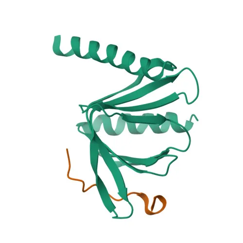
Monoclonal Antibodies
Anti-DCP1A Rabbit Monoclonal Antibody for WB, IHC-P, IF/ICC, ELISA - Q9NPI6
Item Number : CM0011237
Price varies based on specs and customizations
- Application
- WB, IHC-P, IF/ICC, ELISA
- Cross Reactivity
- Human, Mouse
- Protein Weight
- 63kDa

Shipping:
Contact us to get shipping details Enjoy On-time Dispatch Guarantee.
Why Choose Us
Easy ordering process, quality products, and dedicated support for your business success.
Core Product Specifications and Parameters
| Parameter | Value |
|---|---|
| Product Name | DCP1A Rabbit mAb |
| Remarks/Alias | SMIF, SMAD4IP1, HSA275986, Nbla00360, DCP1A |
| Species | Human |
| GeneID (Human) | 55802 |
| Immunogen | A synthetic peptide corresponding to a sequence within amino acids 483-582 of human DCP1A (Q9NPI6). |
| Source | Rabbit |
| Category | Monoclonal Antibodies |
| Application | WB, IHC-P, IF/ICC, ELISA |
| Cross Reactivity | Human, Mouse |
| SWISS | Q9NPI6 |
| Protein Weight | 63kDa |
| Shipping | Ice bag |
Biological Background: DCP1A Function and Localization
- DCP1A (mRNA-decapping enzyme 1A) is a hydrolase that removes the 7-methyl guanine cap from mRNA molecules, generating 5'-phosphorylated mRNA and 7m-GDP, necessary for normal mRNA turnover and nonsense-mediated mRNA decay. Related references: PMID:12417715
- It also functions as a transcriptional co-activator (named SMIF) that contributes to transactivation of target genes after TGFB1 stimulation. Related references: PMID:11836524
- DCP1A is essential for embryonic development. Related references: PMID:33813271
- Localizes to the cytoplasm, specifically in P-bodies, and also in the nucleus.
- Detected in multiple tissues including heart, brain, placenta, lung, skeletal muscle, liver, kidney, and pancreas.
- The protein undergoes post-translational modifications such as phosphorylation and methylation; it is a member of the reference proteome.
- Alternative splicing produces multiple isoforms.
Experimental Guidance and Technical Tips
- The immunogen corresponds to a peptide within amino acids 483–582 (C-terminal region), so the antibody is expected to recognize full-length DCP1A. Confirm specificity by assessing band size and, if needed, knock-down/knock-out validation.
- For Western blot (WB), a band around 63 kDa is expected. Use positive controls from human or mouse cell lines known to express DCP1A.
- For immunohistochemistry (IHC-P), validate the antibody in paraffin-embedded tissue sections with known DCP1A expression, such as heart or placenta.
- For immunofluorescence (IF/ICC), DCP1A typically shows cytoplasmic P-body puncta and may also show nuclear staining; optimize fixation and permeabilization conditions for the cell type used.
- For ELISA, the synthetic peptide immunogen can be used as a positive control to confirm antibody binding.
CamelBio: Your One-Stop Sourcing Bridge
CamelBio offers diagnostic manufacturers and research institutions a streamlined, one-stop sourcing platform for IVD raw materials. For DCP1A and associated mRNA decapping or TGF‑β signaling research, we can supply validated antibody pairs, optimized monoclonal antibodies like this anti-DCP1A clone, bulk ancillary reagents, and bespoke sourcing for rare targets. Partner with CamelBio to efficiently bridge your concept-to-clinic development needs.
Product Datasheet
Anti-DCP1A Rabbit Monoclonal Antibody for WB, IHC-P, IF/ICC, ELISA - Q9NPI6
REQUEST A QUOTE
Our professional team will reply to you within one business day. Please feel free to contact us!