Polyclonal Antibodies
Anti-DAP Kinase 1 (DAPK1) Polyclonal Antibody for WB, IHC-P, ELISA - P53355
Item Number : CM0027435
Price varies based on specs and customizations
- Application
- WB, IHC-P, ELISA
- Cross Reactivity
- Human, Mouse, Rat
- Protein Weight
- 160kDa

Shipping:
Contact us to get shipping details Enjoy On-time Dispatch Guarantee.
Why Choose Us
Easy ordering process, quality products, and dedicated support for your business success.
Core Product Specifications and Parameters
| Parameter | Value |
|---|---|
| Product Name | DAP Kinase 1 (DAPK1) Rabbit pAb |
| Remarks/Alias | DAPK; ROCO3; DAP Kinase 1 (DAPK1) |
| Species | Human |
| Gene ID (Human) | 1612 |
| Gene ID | 1612 |
| Immunogen | Recombinant fusion protein containing a sequence corresponding to amino acids 1141-1430 of human DAP Kinase 1 (DAPK1) (NP_004929.2) |
| Source | Rabbit |
| Category | Polyclonal Antibodies |
| Application | WB, IHC-P, ELISA |
| Cross Reactivity | Human, Mouse, Rat |
| SWISS | P53355 |
| Protein Weight | 160kDa |
| Shipping | Ice bag |
Biological Background: DAP Kinase 1 (DAPK1) Function and Localization
- DAPK1 is a calcium/calmodulin-dependent serine/threonine kinase that integrates multiple cellular signaling pathways, governing cell survival, apoptosis, and autophagy.
- It regulates both caspase-dependent type I apoptotic death and caspase-independent type II autophagic cell death, depending on cellular context.
- DAPK1 phosphorylates a diverse array of substrates—including PIN1, TPM1, STX1A, PRKD1, BECN1, TSC2, RPS6, MYL9, and DAPK3—modulating nuclear localization, cytoskeletal dynamics, vesicle trafficking, and metabolic signaling.
- In stroke, DAPK1 acts as an NMDA receptor signaling amplifier, phosphorylating GRINB at Ser-1303 to drive injurious Ca2+ influx and irreversible neuronal death.
- Isoform 2, generated by alternative splicing, cannot induce apoptosis but promotes membrane blebbing; it is expressed in normal intestinal tissue and colorectal carcinomas.
- Subcellular localization is predominantly cytoplasmic and associated with the cytoskeleton, consistent with its roles in remodeling and signaling.
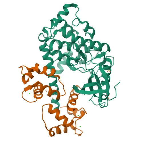
- The protein features a multidomain architecture that includes a kinase domain, a calmodulin-binding domain, ankyrin repeats, and a death domain, underpinning its function in cell death regulation.
Experimental Guidance and Technical Tips
- For WB, a specific band at approximately 160 kDa is expected in lysates from human, mouse, or rat samples; confirm specificity using appropriate positive and negative controls.
- In IHC-P, optimize antigen retrieval and antibody dilution; consider known expression patterns (e.g., brain, intestinal epithelium) for validation.
- For ELISA, the antibody can be used as a capture or detection reagent; empirical determination of coating concentration and pairing partners is recommended.
- Because the immunogen maps to the C-terminal region (aa 1141-1430), verify detection of isoform 2 in relevant experimental systems if required.
CamelBio: Your One-Stop Sourcing Bridge
CamelBio supports diagnostic manufacturers and research laboratories worldwide with a comprehensive portfolio of IVD raw materials, including high-quality antibodies targeting key regulators like DAPK1. Whether you need validated antibody pairs, optimized polyclonal or monoclonal reagents, bulk ancillary products, or assistance sourcing rare-target raw materials for apoptosis and autophagy studies, CamelBio provides end-to-end procurement and technical consultation to accelerate your assay development.
Product Datasheet
Anti-DAP Kinase 1 (DAPK1) Polyclonal Antibody for WB, IHC-P, ELISA - P53355
REQUEST A QUOTE
Our professional team will reply to you within one business day. Please feel free to contact us!