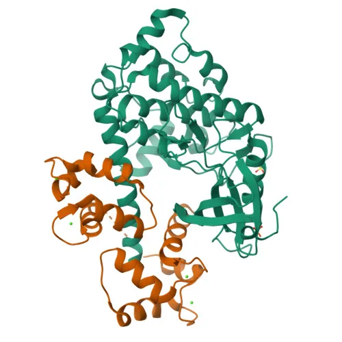
Monoclonal Antibodies
Anti-DAP Kinase 1 (DAPK1) Monoclonal Antibody for WB and ELISA - P53355
Item Number : CM0002464
Price varies based on specs and customizations
- Application
- WB, ELISA
- Cross Reactivity
- Human, Mouse
- Protein Weight
- 160kDa

Shipping:
Contact us to get shipping details Enjoy On-time Dispatch Guarantee.
Why Choose Us
Easy ordering process, quality products, and dedicated support for your business success.
Core Product Specifications and Parameters
| Parameter | Value |
|---|---|
| Product Name | DAP Kinase 1 (DAPK1) Rabbit mAb |
| Remarks/Alias | DAPK, ROCO3, DAP Kinase 1 (DAPK1) |
| Species | Human |
| Gene ID | 1612 |
| Immunogen | Synthetic Peptide |
| Source | Rabbit |
| Category | Monoclonal Antibodies |
| Application | WB, ELISA |
| Cross Reactivity | Human, Mouse |
| SWISS | P53355 |
| Protein Weight | 160kDa |
| Shipping | Ice bag |
Biological Background: DAP Kinase 1 (DAPK1) Function and Localization
- Encodes a calcium/calmodulin-dependent serine/threonine kinase that integrates signals for apoptosis, autophagy, and cell survival depending on cellular context.
- Mediates both caspase-dependent type I apoptotic death and caspase-independent type II autophagic death characterized by autophagic vesicle accumulation.
- Regulates diverse substrates including PIN1, TPM1, STX1A, PRKD1, BECN1, TSC2, RPS6, MYL9, DAPK3, and GRINB, thereby influencing cytoskeletal dynamics, exocytosis, stress signaling, autophagy induction, mTORC1 activity, and NMDA receptor-mediated excitotoxicity.
- Isoform 2 lacks apoptotic activity but retains the ability to induce membrane blebbing; it is expressed in normal intestinal tissue and colorectal carcinomas.
- Localizes to the cytoplasm and associates with the cytoskeleton.
- Contains distinctive domains: N-terminal kinase domain, calmodulin-binding region, ankyrin repeats, and a C-terminal death domain; also possesses ATP- and GTP-binding motifs.
- Listed under keywords including alternative splicing, apoptosis, autophagy, phosphoprotein, and Ubl conjugation, reflecting its complex regulation and functional versatility.
Experimental Guidance and Technical Tips
- The immunogen lies within residues 250–350, a region between the kinase domain and the calmodulin-binding motif; consider verifying epitope accessibility under native versus denaturing conditions.
- For Western blot, a primary band at ~160 kDa represents full-length DAPK1; additional bands may arise from splice isoforms or degradation products—validating with appropriate controls (e.g., knockdown or knockout lysates) is recommended.
- Confirm cross-reactivity in mouse samples, as the antibody is reported to recognize both human and mouse DAPK1.
- For ELISA, standard indirect or sandwich protocols can be applied; optimize coating concentration and detection conditions for your specific assay format.
- Take into account that isoform 2 may not contain the peptide sequence; check expression profiles in your sample set (e.g., normal vs. cancerous intestinal tissue) to avoid misinterpretation.
CamelBio: Your One-Stop Sourcing Bridge
CamelBio provides a streamlined sourcing channel for IVD raw materials, including validated antibodies for key signaling targets such as DAPK1. Our portfolio spans antibody pairs, monoclonal and polyclonal antibodies, bulk ancillary reagents, and custom sourcing for rare targets—supporting laboratories and diagnostic developers from initial concept to clinical translation. We invite you to explore our comprehensive solutions for apoptosis and autophagy research needs.
REQUEST A QUOTE
Our professional team will reply to you within one business day. Please feel free to contact us!