Polyclonal Antibodies
Anti-DAG1 Polyclonal Antibody for WB, IF/ICC, ELISA - Q14118
Item Number : CM0024029
Price varies based on specs and customizations
- Application
- WB, IF/ICC, ELISA
- Cross Reactivity
- Human, Mouse, Rat
- Protein Weight
- 97kDa

Shipping:
Contact us to get shipping details Enjoy On-time Dispatch Guarantee.
Why Choose Us
Easy ordering process, quality products, and dedicated support for your business success.
Core Product Specifications and Parameters
| Parameter | Value |
|---|---|
| Product Name | DAG1 Rabbit pAb |
| Remarks/Alias | A3a; DAG; AGRNR; 156DAG; MDDGA9; MDDGC7; MDDGC9; LGMDR16; DAG1 |
| Species | Human |
| GeneID (human) | 1605 |
| GeneID | 1605 |
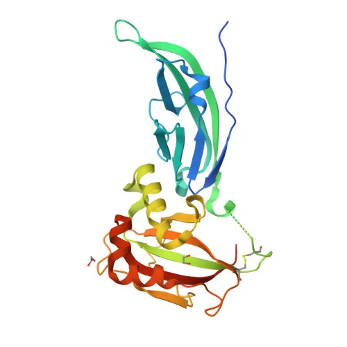
| Immunogen | Recombinant fusion protein containing a sequence corresponding to amino acids 654-749 of human DAG1 (NP_004384.4). |
| Source | Rabbit |
| Category | Polyclonal Antibodies |
| Application | WB, IF/ICC, ELISA |
| Cross Reactivity | Human, Mouse, Rat |
| SWISS | Q14118 |
| Protein Weight | 97kDa |
| Shipping | Ice bag |
Biological Background: DAG1 Function and Localization
- Dystroglycan 1 (DAG1) is a central component of the dystrophin–glycoprotein complex, connecting the extracellular matrix to the intracellular cytoskeleton. It is expressed as a precursor that undergoes autocatalytic cleavage into peripheral alpha‑dystroglycan and transmembrane beta‑dystroglycan.
- The dystroglycan complex participates in laminin deposition and extracellular matrix assembly, acetylcholine receptor clustering at the neuromuscular junction, sarcolemmal stability, cell survival, peripheral nerve myelination, cell migration, epithelial polarization, and epithelial branching morphogenesis.
- Alpha‑dystroglycan binds laminin‑G‑domain‑containing extracellular matrix proteins (e.g., laminin‑1/‑2, agrin, perlecan) and mediates signaling via PI3K/AKT and Src‑kinase pathways, thereby regulating apoptotic signals and synaptic organization.
- Beta‑dystroglycan anchors to dystrophin (DMD) or utrophin (UTRN) and scaffolds axin to the actin cytoskeleton, facilitating cell adhesion‑mediated signaling and cell polarity.
- In cartilage, DAG1 binds agrin and up‑regulates the chondrogenic transcription factor SOX9, positively regulating cartilage formation.
Related references: PMID:26290588 - The protein localizes to multiple compartments: the sarcolemma and postsynaptic membrane, synaptic sites, the basement membrane, extracellular space, cytoplasm/cytoskeleton, and the nucleoplasm.
- DAG1 acts as a host cell receptor for class‑C new‑world arenaviruses (Lassa, lymphocytic choriomeningitis virus) and for Mycobacterium leprae entry into Schwann cells.
Related references: PMID:16254364, PMID:17360738, PMID:19324387, PMID:9851927 - Expressed in a variety of fetal and adult tissues, with prominent localization to the basement membrane in epidermis and expression in keratinocytes and fibroblasts.
Experimental Guidance and Technical Tips
- The immunogen (aa 654–749) resides in the C‑terminal portion of beta‑dystroglycan; the antibody is expected to recognize both the full‑length ~97 kDa precursor and the cleaved beta‑subunit (~43 kDa) in denaturing/reducing Western blots.
- For Western blotting, validate the observed band pattern in known positive tissues/cell lines (e.g., skeletal muscle, brain) and consider using fresh protease‑inhibited lysates to prevent degradation.
- In immunofluorescence/ICC, permeabilization is required to access the intracellular epitope. Co‑staining with sarcolemmal or basement membrane markers can confirm localization.
- For ELISA applications, titrate the antibody as capture or detection reagent using recombinant DAG1 fragments or cell lysates under standard conditions; performance should be optimized in the end‑user’s assay system.
- Species cross‑reactivity has been reported for human, mouse, and rat; if working with other species, test experimentally and confirm sequence homology beforehand.
CamelBio: Your One-Stop Sourcing Bridge
CamelBio provides diagnostic manufacturers and research laboratories with a streamlined, one‑stop channel for IVD raw materials. This anti‑DAG1 polyclonal antibody supports investigation of muscular dystrophies, cell adhesion processes, and viral pathogenesis. Beyond individual antibodies, CamelBio offers validated antibody pairs, bulk ancillary reagents, and specialized sourcing for rare‑target raw materials to accelerate your assay development from concept to clinic.
REQUEST A QUOTE
Our professional team will reply to you within one business day. Please feel free to contact us!