Monoclonal Antibodies
Anti-Cytokeratin 19 (CK19) Rabbit Monoclonal Antibody for WB, IF/ICC, IF-P, IHC-P, ELISA - P08727
Item Number : CM0003350
Price varies based on specs and customizations
- Application
- WB, IF/ICC, IF-P, IHC-P, ELISA
- Cross Reactivity
- Human
- Protein Weight
- 44 kDa

Shipping:
Contact us to get shipping details Enjoy On-time Dispatch Guarantee.
Why Choose Us
Easy ordering process, quality products, and dedicated support for your business success.
Core Product Specifications and Parameters
| Parameter | Specification |
|---|---|
| Product Name | Cytokeratin 19 (CK19) Rabbit mAb |
| Remarks/Alias | K19; KRT19; K1CS; Cytokeratin 19 (CK19) |
| Species | Human |
| GeneID (human) | 3880 |
| GeneID | 3880 |
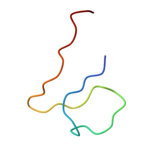
| Immunogen | Synthetic Peptide|A synthetic peptide corresponding to a sequence within amino acids 1-100 of human Cytokeratin 19 (KRT19) (KRT19) (P08727). |
| Source | Rabbit |
| Category | Monoclonal Antibodies |
| Application | WB, IF/ICC, IF-P, IHC-P, ELISA |
| Cross Reactivity | Human |
| SWISS | P08727 |
| Protein Weight | 44 kDa |
| Shipping | Ice bag |
Biological Background: Cytokeratin 19 (CK19) Function and Localization
- Cytokeratin 19 (CK19, K19) is a type I keratin encoded by the KRT19 gene. It is an intermediate filament protein with a molecular weight of approximately 44 kDa.
- CK19, along with its binding partner KRT8 (type II keratin), is involved in the organization of myofibers. Together they help link the contractile apparatus to dystrophin at the costameres of striated muscle.
- CK19 is expressed in a wide range of epithelial tissues, including basal keratinocytes of the hair follicle outer root sheath, sweat gland and mammary gland ductal and secretory cells, bile ducts, gastrointestinal tract, bladder urothelium, oral epithelia, esophagus, and ectocervical epithelium. It is also found in a subset of vascular wall cells and in muscle fibers accumulating at costameres.
- Subcellularly, CK19 localizes to cytoplasmic intermediate filaments and at costameres in striated muscle, associating with dystrophin and spectrin at the sarcolemma.
- Post-translational modifications include phosphorylation and methylation, which may regulate filament dynamics.
- CK19 is annotated to interact with viral proteins (host-virus interaction keyword), suggesting a role in viral infection processes.
- Structural studies reveal a coiled-coil domain characteristic of keratins, and the protein has been directly sequenced and identified by proteomics (reference proteome).
Experimental Guidance and Technical Tips
- The immunogen corresponds to amino acids 1-100 in the N-terminal region of human CK19. This region may be conserved across isoforms; consider sequence alignment to verify specificity if using other species.
- For western blotting, a band at ~44 kDa is expected. Confirm appropriate loading controls and optimize antibody dilution for your sample type.
- In immunofluorescence and immunohistochemistry, CK19 exhibits cytoplasmic filamentous staining in epithelial cells and costameric localization in muscle. Validate staining patterns in positive control tissues such as colon, breast, or skin.
- For ELISA applications, consider using recombinant CK19 protein or cell lysates to determine optimal coating conditions and detection range.
- Always include appropriate negative controls (e.g., knockout cell lines or tissues known to lack CK19 expression) to confirm specificity.
CamelBio: Your One-Stop Sourcing Bridge
CamelBio offers diagnostic developers and research laboratories streamlined access to high-quality IVD raw materials. This anti-CK19 rabbit monoclonal antibody exemplifies our portfolio of optimized antibodies for epithelial and cancer biomarker research. Beyond individual antibodies, we supply validated antibody pairs, bulk ancillary reagents, and support for rare-target sourcing, helping you bridge from concept to clinic with confidence.
Product Datasheet
Anti-Cytokeratin 19 (CK19) Rabbit Monoclonal Antibody for WB, IF/ICC, IF-P, IHC-P, ELISA - P08727
REQUEST A QUOTE
Our professional team will reply to you within one business day. Please feel free to contact us!