Polyclonal Antibodies
Anti-Cytohesin-1 (CYTH1) Polyclonal Antibody for WB, IF/ICC, ELISA - Q15438
Item Number : CM0021032
Price varies based on specs and customizations
- Application
- WB, IF/ICC, ELISA
- Cross Reactivity
- Human, Mouse, Rat
- Protein Weight
- 46kDa

Shipping:
Contact us to get shipping details Enjoy On-time Dispatch Guarantee.
Why Choose Us
Easy ordering process, quality products, and dedicated support for your business success.
Core Product Specifications and Parameters
| Parameter | Value |
|---|---|
| Product Name | CYTH1 Rabbit pAb |
| Remarks/Alias | B2-1; SEC7; PSCD1; D17S811E; CYTOHESIN-1; CYTH1 |
| Species | Human |
| GeneID (Human) | 9267 |
| Immunogen | Recombinant fusion protein containing a sequence corresponding to amino acids 1-70 of human CYTH1 (NP_004753.1) |
| Source | Rabbit |
| Category | Polyclonal Antibodies |
| Application | WB, IF/ICC, ELISA |
| Cross Reactivity | Human, Mouse, Rat |
| SWISS | Q15438 |
| Protein Weight | 46kDa |
| Shipping | Ice bag |
Biological Background: Cytohesin-1 Function and Localization
- Cytohesin-1 (CYTH1) is a member of the cytohesin family of guanine nucleotide exchange factors (GEFs) that activate ADP-ribosylation factor (ARF) small GTPases, serving as a crucial regulator of intracellular vesicular trafficking.
- The protein is characterized by a pleckstrin homology (PH) domain for phosphoinositide binding, a Sec7 domain responsible for its GEF activity, and a coiled-coil domain involved in protein-protein interactions.
- CYTH1 specifically catalyzes guanine-nucleotide exchange on ARF1, ARF5, and ARF6 by promoting the replacement of GDP with GTP, thereby controlling their activation state.
- Through ARF6 regulation, it plays a pivotal role in membrane trafficking events essential for junctional remodeling and the establishment of epithelial cell polarity.
- Ubiquitously expressed across tissues, reflecting its fundamental role in cellular transport processes.
- Subcellularly, CYTH1 localizes to the cell membrane, cytosol, and cell-cell junctions including tight junctions and adherens junctions, consistent with its functions in membrane dynamics and cell adhesion.
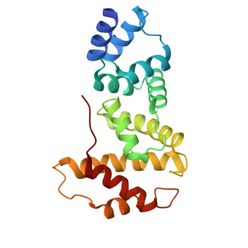
- Post-translational modifications include acetylation and ubiquitin conjugation, and the protein's three-dimensional structure has been resolved.
Experimental Guidance and Technical Tips
- The immunogen consists of the N-terminal 70 amino acids of human CYTH1, a region that may be conserved across species (supporting the antibody's cross-reactivity with mouse and rat) and potentially retained in alternative splicing isoforms.
- For Western blotting, the expected band migrates at approximately 46 kDa; optimal antibody dilution and blocking conditions should be empirically determined using positive control lysates (e.g., HeLa, HEK293, or U-2 OS cells).
- In immunofluorescence/immunocytochemistry, fixatives such as 4% paraformaldehyde and permeabilization with 0.1–0.3% Triton X-100 are recommended; however, the chosen method should preserve membrane and junctional structures where CYTH1 localizes.
- For ELISA applications, the antibody may be employed as either a capture or detection reagent after appropriate optimization, including testing different coating and blocking conditions.
CamelBio: Your One-Stop Sourcing Bridge
CamelBio simplifies IVD raw-material procurement for diagnostic manufacturers and research laboratories. The CYTH1 polyclonal antibody is just one example from our extensive portfolio supporting ARF signaling, membrane trafficking, and epithelial biology research. We offer validated antibody pairs, optimized monoclonal/polyclonal antibodies, bulk ancillary reagents, and custom sourcing for rare targets—all backed by technical consulting to help you move from concept to clinic seamlessly.
Product Datasheet
Anti-Cytohesin-1 (CYTH1) Polyclonal Antibody for WB, IF/ICC, ELISA - Q15438
REQUEST A QUOTE
Our professional team will reply to you within one business day. Please feel free to contact us!