Monoclonal Antibodies
Anti-Cystatin C Rabbit Monoclonal Antibody for WB, ELISA - P01034
Item Number : CM0012586
Price varies based on specs and customizations
- Application
- WB, ELISA
- Cross Reactivity
- Human, Mouse
- Protein Weight
- 13kDa

Shipping:
Contact us to get shipping details Enjoy On-time Dispatch Guarantee.
Why Choose Us
Easy ordering process, quality products, and dedicated support for your business success.
Core Product Specifications and Parameters
| Parameter | Value |
|---|---|
| Product Name | Cystatin C Rabbit mAb |
| Remarks/Alias | ARMD11; HEL-S-2; Cystatin C |
| Species | Human |
| Gene ID (Human) | 1471 |
| Gene ID | 1471 |
| Immunogen | Recombinant fusion protein containing a sequence corresponding to amino acids 1-146 of human Cystatin C (P01034) |
| Source | Rabbit |
| Category | Monoclonal Antibodies |
| Application | WB, ELISA |
| Cross Reactivity | Human, Mouse |
| SWISS | P01034 |
| Protein Weight | 13kDa |
| Shipping | Ice bag |
Biological Background: Cystatin C Function and Localization
- Cystatin C (also known as cystatin-3, gamma-trace, neuroendocrine basic polypeptide, post-gamma-globulin) is a secreted cysteine protease inhibitor encoded by the CST3 gene.
- It functions as a potent inhibitor of lysosomal cysteine proteinases (cathepsins B, H, L, S) and extracellular papain-like enzymes, regulating proteolytic activity in various biological fluids.
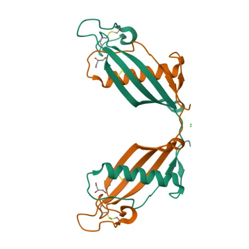
- Belongs to the cystatin superfamily (type 2 cystatins), sharing a conserved cystatin fold with disulfide bonds critical for structural stability.
- Predominantly localized to the extracellular space; it is secreted and found in high concentrations in cerebrospinal fluid, plasma, and other body fluids.
- Exhibits tissue-specific expression: highest in epididymis, vas deferens, brain, thymus, and ovary; lower in submandibular gland; present in submandibular and sublingual saliva but absent in parotid saliva.
- Post-translational modifications include signal peptide cleavage, N-linked glycosylation, and phosphorylation, contributing to its functional diversity.
- Implicated in multiple pathologies, including age-related macular degeneration (ARMD11), cerebral amyloid angiopathy, hereditary cystatin C amyloid angiopathy (HCCAA), and leukodystrophy, often involving amyloid fibril formation.
- Diagnostic significance: serum cystatin C levels are used as a marker of kidney function, and its presence in body fluids aids in monitoring renal and neurological disorders.
Experimental Guidance and Technical Tips
- For WB, consider using a positive control such as human serum or cerebrospinal fluid, where cystatin C is abundant; expect a band around 13 kDa for the mature protein under reducing conditions.
- When probing mouse samples, validate cross-reactivity initially, as the antibody detects both human and mouse cystatin C; mouse kidney or serum can serve as positive controls.
- For ELISA, pairing this capture antibody with a detection antibody of different clonality or epitope may enhance signal; optimize coating and blocking conditions for native secreted cystatin C.
- Ensure thorough sample preparation for low-molecular-weight proteins (≤15 kDa) by using appropriate membrane pore sizes and transfer times to avoid loss during western blotting.
- Consider using recombinant human cystatin C as a standard and positive control for quantitative assays.
CamelBio: Your One-Stop Sourcing Bridge
CamelBio provides diagnostic manufacturers, labs, and research institutes with one-stop access to validated IVD raw materials, including optimized monoclonal antibodies like this cystatin C clone, bulk ancillary reagents, and technical support for assay development. This antibody accelerates research on renal function biomarkers and neurodegenerative disease targets, from concept to clinical application. For rare target sourcing or custom antibody services, CamelBio’s expertise ensures seamless integration into your diagnostic pipeline.
REQUEST A QUOTE
Our professional team will reply to you within one business day. Please feel free to contact us!