Monoclonal Antibodies
Anti-Cystatin A Monoclonal Antibody for WB, IHC-P, IF/ICC, ELISA - P01040
Item Number : CM0010107
Price varies based on specs and customizations
- Application
- WB, IHC-P, IF/ICC, ELISA
- Cross Reactivity
- Human, Mouse, Rat
- Protein Weight
- 11kDa

Shipping:
Contact us to get shipping details Enjoy On-time Dispatch Guarantee.
Why Choose Us
Easy ordering process, quality products, and dedicated support for your business success.
Core Product Specifications and Parameters
| Parameter | Value |
|---|---|
| Product Name | Cystatin A Rabbit mAb |
| Remarks/Alias | AREI; PSS4; STF1; STFA; Cystatin A |
| Species | Human |
| Gene ID (Human) | 1475 |
| Gene ID | 1475 |
| Immunogen | Synthetic Peptide|A synthetic peptide corresponding to a sequence within amino acids 1-98 of human Cystatin A (NP_005204.1). |
| Source | Rabbit |
| Category | Monoclonal Antibodies |
| Application | WB, IHC-P, IF/ICC, ELISA |
| Cross Reactivity | Human, Mouse, Rat |
| SWISS | P01040 |
| Protein Weight | 11kDa |
| Shipping | Ice bag |
Biological Background: Cystatin A Function and Localization
- Cystatin A, also known as Stefin-A or Cystatin-AS, is encoded by the CSTA gene (aliases STF1, STFA) and belongs to the cystatin superfamily of cysteine protease inhibitors (UniProt P01040).
- It functions primarily as an intracellular thiol (cysteine) protease inhibitor, regulating proteolytic activity within the cytoplasm.
- Beyond its inhibitory role, Cystatin A contributes to desmosome-mediated cell-cell adhesion, particularly in the lower layers of the epidermis.
- Subcellularly, Cystatin A is localized to the cytoplasm.
- Expression of Cystatin A is prominent in the skin, where it is found throughout the epidermis.
- The protein has a calculated molecular weight of approximately 11 kDa (11,006 Da) and undergoes post-translational modifications including N-terminal acetylation.
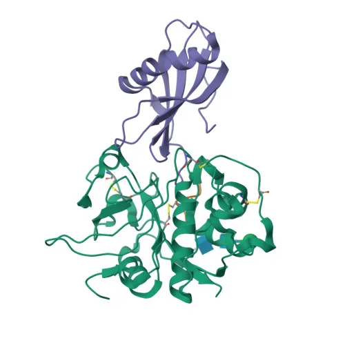
- Clinically, Cystatin A is associated with ichthyosis, and its crystal structure has been solved, aiding functional studies.
Experimental Guidance and Technical Tips
- The immunogen used to generate this antibody corresponds to the N-terminal region (aa 1-98) of full-length human Cystatin A. Researchers may consider this region's conservation when predicting cross-reactivity with orthologs from other species (confirmed for mouse and rat).
- This antibody is validated for Western blotting (WB), immunohistochemistry on paraffin-embedded sections (IHC-P), immunofluorescence/immunocytochemistry (IF/ICC), and ELISA. For WB, given the small size of Cystatin A (~11 kDa), ensure appropriate gel percentage and membrane transfer conditions to resolve the protein. For IHC-P and IF/ICC, optimize antigen retrieval and permeabilization steps, as Cystatin A is intracellular. Always include appropriate positive (e.g., skin tissue lysate) and negative controls.
CamelBio: Your One-Stop Sourcing Bridge
CamelBio serves as a trusted one-stop source for IVD raw materials, supporting diagnostic manufacturers and research laboratories with validated antibodies, antigen pairs, and ancillary reagents. This Cystatin A monoclonal antibody is ideal for developing assays targeting protease inhibition and skin barrier function. Contact us for bulk orders, custom antibody development, or assistance in sourcing rare targets to accelerate your diagnostic and research workflows.
Product Datasheet
Anti-Cystatin A Monoclonal Antibody for WB, IHC-P, IF/ICC, ELISA - P01040
REQUEST A QUOTE
Our professional team will reply to you within one business day. Please feel free to contact us!