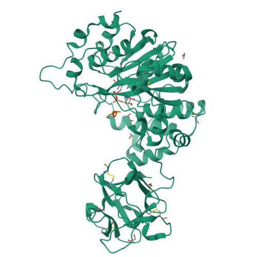
Polyclonal Antibodies
Anti-CYR61 Polyclonal Antibody for WB, IF/ICC, ELISA - O00622
Item Number : CM0017733
Price varies based on specs and customizations
- Application
- WB, IF/ICC, ELISA
- Cross Reactivity
- Human, Mouse, Rat
- Protein Weight
- 42kDa

Shipping:
Contact us to get shipping details Enjoy On-time Dispatch Guarantee.
Why Choose Us
Easy ordering process, quality products, and dedicated support for your business success.
Core Product Specifications and Parameters
| Parameter | Value |
|---|---|
| Product Name | CYR61 Rabbit pAb |
| Remarks/Alias | GIG1; CYR61; IGFBP10 |
| Species | Human |
| GeneID (Human) | 3491 |
| GeneID | 3491 |
| Immunogen | Synthetic peptide|A synthetic peptide corresponding to a sequence within amino acids 120-220 of human CYR61 (NP_001545.2). |
| Source | Rabbit |
| Category | Polyclonal Antibodies |
| Application | WB, IF/ICC, ELISA |
| Cross Reactivity | Human, Mouse, Rat |
| SWISS | O00622 |
| Protein Weight | 42kDa |
| Shipping | Ice bag |
Biological Background: CYR61 Function and Localization
- CYR61, also known as CCN family member 1, cellular communication network factor 1, cysteine-rich angiogenic inducer 61, protein GIG1, and insulin-like growth factor-binding protein 10 (IGFBP-10), is a secreted, extracellular matrix-associated signaling protein.
- It promotes cell proliferation, chemotaxis, angiogenesis, and cell adhesion, playing a key role in wound healing by up-regulating genes involved in angiogenesis, inflammation, and matrix remodeling (e.g., VEGA-A, VEGA-C, MMP1, MMP3, TIMP1, uPA, PAI-1, and integrins alpha-3 and alpha-5) in skin fibroblasts.
- CYR61-mediated gene regulation is dependent on heparin-binding, and it down-regulates the expression of alpha-1 and alpha-2 subunits of collagen type-1.
- It mediates cell adhesion and adhesive signaling through integrin alpha-6/beta-1, cell migration through integrin alpha-v/beta-5, and cell proliferation through integrin alpha-v/beta-3.
- The protein contains key structural features including disulfide bonds and phosphorylation sites, and it carries a signal peptide for secretion, localizing it to the extracellular space.
- In skin fibroblasts, CYR61 functions during wound healing by modulating integrin expression and matrix remodeling.
Experimental Guidance and Technical Tips
- This polyclonal antibody was raised against a synthetic peptide corresponding to amino acids 120–220 of human CYR61; consider using full-length recombinant protein or native samples for specificity validation.
- For Western blotting, a band around 42kDa is expected; due to the secreted nature of CYR61, analyze conditioned medium or cell lysates with proper sample preparation.
- In immunofluorescence/ICC, include a negative control (e.g., peptide blocking) to confirm signal specificity, and optimize permeabilization for intracellular detection despite its secretory pathway.
- ELISA performance will depend on coating conditions; pairing with a detection antibody recognizing a distinct epitope is recommended for sandwich ELISA.
- Cross-reactivity with mouse and rat CYR61 is stated; validate in the relevant sample system for your application.
CamelBio: Your One-Stop Sourcing Bridge
CamelBio provides diagnostic manufacturers, laboratories, and research institutions with integrated access to IVD raw materials, technical services, and consulting—from concept to clinic. For studies on cell adhesion, angiogenesis, and wound healing, our portfolio includes validated antibody pairs, optimized monoclonal/polyclonal antibodies, bulk ancillary reagents, and custom sourcing of rare-target raw materials. Whether you need CYR61 detection reagents or broader CCN family tools, CamelBio serves as your reliable B2B supply partner.
REQUEST A QUOTE
Our professional team will reply to you within one business day. Please feel free to contact us!