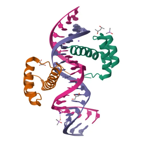
Polyclonal Antibodies
Anti-CRX Rabbit Polyclonal Antibody for WB, IF-P, ELISA - O43186
Item Number : CM0024761
Price varies based on specs and customizations
- Application
- WB, IF-P, ELISA
- Cross Reactivity
- Mouse
- Protein Weight
- 32kDa

Shipping:
Contact us to get shipping details Enjoy On-time Dispatch Guarantee.
Why Choose Us
Easy ordering process, quality products, and dedicated support for your business success.
Core Product Specifications and Parameters
| Parameter | Value |
|---|---|
| Product Name | CRX Rabbit pAb |
| Remarks/Alias | CRD; LCA7; OTX3; CORD2; CRX |
| Species | Human |
| Gene ID (Human) | 1406 |
| Gene ID | 1406 |
| Immunogen | Synthetic peptide: A synthetic peptide corresponding to a sequence within amino acids 100-200 of human CRX (NP_000545.1). |
| Source | Rabbit |
| Category | Polyclonal Antibodies |
| Application | WB, IF-P, ELISA |
| Cross Reactivity | Mouse |
| SWISS | O43186 |
| Protein Weight | 32kDa |
| Shipping | Ice bag |
Biological Background: CRX (Cone-rod homeobox protein) Function and Localization
- Cone-rod homeobox protein (CRX) is a member of the homeobox transcription factor family that plays a critical role in photoreceptor development and maintenance.
- CRX specifically binds and transactivates the consensus sequence 5'-TAATC[CA]-3' located in the promoters of several photoreceptor-specific genes, including opsins.
- It acts synergistically with other transcription factors such as NRL, RORB, and RAX to precisely regulate gene expression programs in photoreceptor cells.
- CRX is essential for the maintenance of mammalian photoreceptors, highlighting its importance in visual function.
- The protein localizes to the nucleus, consistent with its role as a transcription factor.
- Tissue expression of CRX is highly specific to the retina, where it orchestrates retinal cell differentiation.
- Mutations in the CRX gene are associated with inherited retinal dystrophies including cone-rod dystrophy, Leber congenital amaurosis, and retinitis pigmentosa.
Experimental Guidance and Technical Tips
- The immunogen design, spanning amino acids 100-200 of human CRX, targets a region that may be accessible for antibody binding in native protein. Consider validating antibody specificity in your experimental system by testing with known positive controls such as retinal tissue or cell lines.
- For Western blot (WB), the predicted molecular weight of CRX is approximately 32 kDa; a band near this size may be observed when using retinal extracts or CRX-expressing cells. Validate detection conditions in your own sample system.
- Immunofluorescence on paraffin-embedded sections (IF-P) may be suitable for detecting nuclear CRX localization in retinal tissues. Optimize antigen retrieval and antibody dilution for your specific samples.
- Cross-reactivity with mouse is reported; if using mouse models, verify reactivity and specificity in mouse retinal samples through appropriate controls.
- ELISA may be used for quantitative detection; optimize coating and detection conditions according to your assay requirements.
CamelBio: Your One-Stop Sourcing Bridge
CamelBio supports diagnostic manufacturers and research laboratories with integrated access to IVD raw materials and technical services—from early-stage development through clinical applications. For researchers studying retinal transcription factors and vision-related disorders, CamelBio offers validated antibodies, optimized recombinant proteins, and ancillary reagents to accelerate assay development. Whether you need bulk polyclonal antibodies, monoclonal pairs, or rare target reagents, CamelBio provides efficient sourcing solutions tailored to your project requirements.
REQUEST A QUOTE
Our professional team will reply to you within one business day. Please feel free to contact us!