Polyclonal Antibodies
Anti-COPZ1 Polyclonal Antibody for WB, IF, ELISA - P61923
Item Number : CM0013431
Price varies based on specs and customizations
- Application
- WB, IF/ICC, ELISA
- Cross Reactivity
- Human, Mouse
- Protein Weight
- 20kDa

Shipping:
Contact us to get shipping details Enjoy On-time Dispatch Guarantee.
Why Choose Us
Easy ordering process, quality products, and dedicated support for your business success.
Core Product Specifications and Parameters
| Parameter | Specifications |
|---|---|
| Product Name | COPZ1 Rabbit pAb |
| Remarks/Alias | COPZ; CGI-120; HSPC181; zeta-COP; zeta1-COP; COPZ1 |
| Species | Human |
| GeneID (Human) | 22818 |
| GeneID | 22818 |
| Immunogen | Recombinant protein |
| Source | Rabbit |
| Category | Polyclonal Antibodies |
| Application | WB,IF/ICC, ELISA |
| Cross Reactivity | Human, Mouse |
| SWISS | P61923 |
| Protein Weight | 20kDa |
| Shipping | Ice bag |
Biological Background: COPZ1 Function and Localization
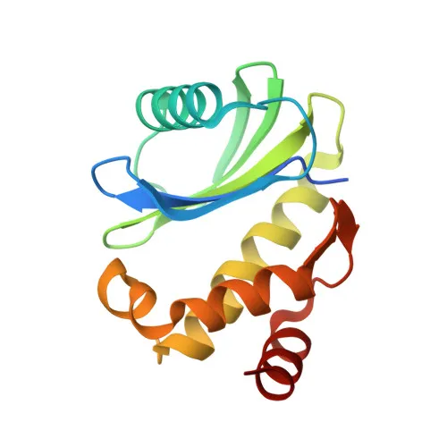
- COPZ1 encodes the coatomer subunit zeta-1, a ~20 kDa component of the COPI coatomer complex essential for intracellular membrane trafficking between the Golgi apparatus and the endoplasmic reticulum (ER).
- It participates directly in retrograde Golgi-to-ER transport by recognizing dilysine motifs on cargo proteins, a process crucial for retrieving escaped ER-resident proteins (PubMed:39642330). Related references: PMID:39642330
- The coatomer complex, including COPZ1, regulates the rate of biosynthetic protein transport through its dynamic association and dissociation with vesicle coats, thereby controlling the budding and cargo selection of COPI-coated vesicles.
- COPZ1 localizes to the cytoplasm, Golgi apparatus membrane, and COPI-coated vesicle membranes, consistent with its role in vesicle formation and cargo selection at the Golgi-ER interface.
- Post-translational acetylation and alternative splicing generate multiple isoforms of COPZ1, which may modulate its activity, stability, or interaction with other coatomer subunits.
- The protein is annotated with the keyword "Disease variant" in UniProt, indicating its potential involvement in human pathologies associated with membrane trafficking defects, although specific disease associations require further experimental validation.
Experimental Guidance and Technical Tips
- The immunogen corresponds to amino acids 1–177 of human COPZ1 (NP_057141.1), which covers the N-terminal region; thus, the polyclonal antibody is expected to recognize full-length and potentially N-terminal truncated isoforms in human and mouse samples.
- For Western blotting, a band around 20 kDa is predicted; we recommend using a reducing loading buffer and validate in relevant cell lines (e.g., HeLa, HEK293, or mouse fibroblasts) to confirm specificity.
- In immunofluorescence/immunocytochemistry (IF/ICC), anticipate a Golgi-like perinuclear staining pattern; co-staining with a Golgi marker (e.g., GM130) can help verify localization.
- ELISA performance should be validated using the recombinant immunogen as a positive control, and optimal antibody dilution must be determined empirically in your assay system.
- Pre-adsorption with the immunogen or use of a blocking peptide (if available) may be employed to confirm signal specificity in critical applications.
CamelBio: Your One-Stop Sourcing Bridge
CamelBio provides IVD manufacturers and research laboratories with streamlined access to high-quality raw materials, leveraging a wide portfolio of validated antibodies, recombinant proteins, and ancillary reagents for diagnostic assay development. For targets like COPZ1, which intersects membrane trafficking and disease-relevant pathways, CamelBio can source rare-target raw materials and offer technical consultation to accelerate your assay optimization. Our integrated supply chain and B2B commitment support you from concept to clinical application, ensuring dependable, scalable material sourcing for your critical workflows.
REQUEST A QUOTE
Our professional team will reply to you within one business day. Please feel free to contact us!