Monoclonal Antibodies
Anti-COPS3 Rabbit Monoclonal Antibody for WB, IHC-P, ELISA - Q9UNS2
Item Number : CM0002684
Price varies based on specs and customizations
- Application
- WB, IHC-P, ELISA
- Cross Reactivity
- Human, Mouse, Rat
- Protein Weight
- 48kDa

Shipping:
Contact us to get shipping details Enjoy On-time Dispatch Guarantee.
Why Choose Us
Easy ordering process, quality products, and dedicated support for your business success.
Core Product Specifications and Parameters
| Parameter | Value |
|---|---|
| Product Name | COPS3 Rabbit mAb |
| Remarks/Alias | CSN3; SGN3; COPS3 |
| Species | Human |
| GeneID (Human) | 8533 |
| GeneID | 8533 |
| Immunogen | Recombinant fusion protein containing a sequence corresponding to amino acids 320-423 of human COPS3 (Q9UNS2) |
| Source | Rabbit |
| Category | Monoclonal Antibodies |
| Application | WB, IHC-P, ELISA |
| Cross Reactivity | Human, Mouse, Rat |
| SWISS | Q9UNS2 |
| Protein Weight | 48kDa |
| Shipping | Ice bag |
Biological Background: COPS3 Function and Localization
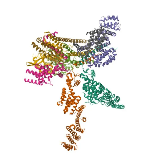
- COPS3, also known as CSN3 or JAB1-containing signalosome subunit 3, is a core component of the COP9 signalosome (CSN) complex.
- The CSN complex is a key regulator of the ubiquitin‑proteasome system, primarily mediating deneddylation of cullin subunits of SCF‑type E3 ubiquitin ligases, which reduces their ligase activity.
- The CSN also participates in phosphorylation of critical signaling proteins, including p53/TP53, c‑jun/JUN, and IκBα/NFKBIA, thereby influencing their stability and degradation.
- COPS3 specifically mediates the interaction between the desmosome and the CSN complex, and this interaction promotes keratinocyte differentiation via deneddylation of EGFR, leading to reduced EGFR stabilization and membrane localization. Related references: PMID:28891468
- Subcellularly, COPS3 is found in the cytoplasm, nucleus, and at cell junctions, reflecting its dynamic roles in signal transduction and adhesion.
- It is widely expressed, with the highest levels observed in heart and skeletal muscle.
- Post‑translational modifications include acetylation and phosphorylation, and alternative splicing generates multiple protein isoforms.
Experimental Guidance and Technical Tips
- The immunogen corresponds to the C‑terminal region (aa 320–423) of human COPS3; epitopes recognized by the antibody likely reside within this fragment.
- For Western blot, a single band at approximately 48 kDa is expected in human, mouse, and rat lysates. Include appropriate positive controls (e.g., HeLa or HEK293 cell lysate) and validate the observed band size in your sample system.
- For immunohistochemistry (paraffin‑embedded sections, IHC‑P), antigen retrieval may be necessary due to the nature of the epitope. Optimize retrieval conditions and antibody dilution using tissue sections known to express COPS3, such as heart or skeletal muscle.
- In ELISA applications, consider using a recombinant COPS3 protein or peptide as the coating antigen to confirm specificity. Titrate the antibody to determine an appropriate working concentration for your assay format.
CamelBio: Your One-Stop Sourcing Bridge
CamelBio offers a comprehensive portfolio of IVD raw materials, including validated antibodies for signalosome and ubiquitin pathway targets like COPS3. Beyond individual antibodies, we supply optimized antibody pairs, monoclonal/polyclonal reagents, bulk ancillary materials, and can assist with sourcing rare or custom raw materials. Backed by end‑to‑end technical support, CamelBio streamlines your path from research to diagnostic development.
REQUEST A QUOTE
Our professional team will reply to you within one business day. Please feel free to contact us!