Polyclonal Antibodies
Anti-COPG1 Rabbit Polyclonal Antibody for WB, IF/ICC, ELISA - Q9Y678
Item Number : CM0017689
Price varies based on specs and customizations
- Application
- WB, IF/ICC, ELISA
- Cross Reactivity
- Human, Mouse, Rat
- Protein Weight
- 98kDa

Shipping:
Contact us to get shipping details Enjoy On-time Dispatch Guarantee.
Why Choose Us
Easy ordering process, quality products, and dedicated support for your business success.
Core Product Specifications and Parameters
| Parameter | Value |
|---|---|
| Product Name | COPG1 Rabbit pAb |
| Remarks/Alias | COPG; COPG1 |
| Species | Human |
| Gene ID (Human) | 22820 |
| Gene ID | 22820 |
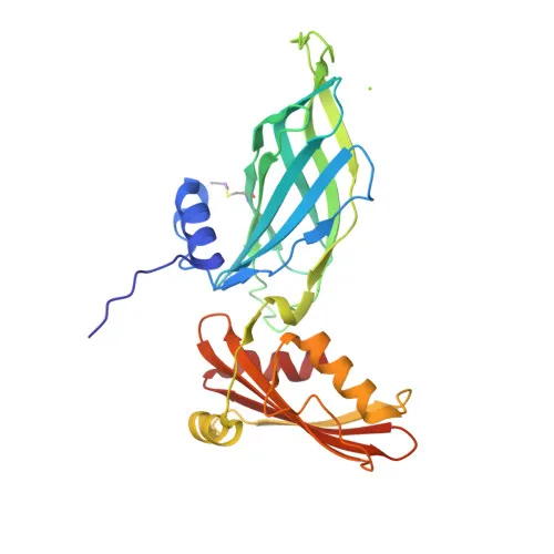
| Immunogen | Recombinant fusion protein containing a sequence corresponding to amino acids 555-874 of human COPG1 (NP_057212.1) |
| Source | Rabbit |
| Category | Polyclonal Antibodies |
| Application | WB, IF/ICC, ELISA |
| Cross Reactivity | Human, Mouse, Rat |
| SWISS | Q9Y678 |
| Protein Weight | 98kDa |
| Shipping | Ice bag |
Biological Background: COPG1 Function and Localization
- COPG1 encodes coatomer subunit gamma-1, a protein component of the COPI vesicle coat complex.
- The coatomer complex reversibly associates with Golgi non-clathrin-coated vesicles and binds dilysine motifs, mediating biosynthetic protein transport and retrograde Golgi-to-ER retrieval of escaped ER residents.
- Coatomer is required for budding from Golgi membranes, influencing Golgi structural integrity, LDL receptor processing and endocytic recycling.
- COPG1 participates in lipid homeostasis by limiting lipid droplet storage; it regulates perilipin family members PLIN2 and PLIN3 localization and promotes PNPLA2 (adipocyte triglyceride lipase) recruitment for lipolysis.
- Subcellular localization includes cytoplasm, Golgi apparatus membrane, and COPI-coated vesicle membrane.
- Post-translational modifications include phosphorylation; disease variants are annotated (UniProt keyword).
Experimental Guidance and Technical Tips
- The immunogen is a recombinant fragment covering amino acids 555–874; the antibody likely detects full-length endogenous COPG1 at ~98 kDa in WB. Confirm specificity using positive and negative controls.
- For WB, consider using treated vs. untreated cell lysates (e.g., HeLa, NIH/3T3) to verify band size and upregulation under Golgi stress or lipolytic conditions.
- For IF/ICC, permeabilization is essential due to cytoplasmic and membrane-associated target localization; co-stain with a Golgi marker to validate.
- In ELISA, assess coating antigen format (peptide vs. recombinant protein) to ensure proper antibody capture.
CamelBio: Your One-Stop Sourcing Bridge
CamelBio provides diagnostic manufacturers, labs, and research institutes with one-stop access to IVD raw materials, technical services, and consulting—covering every stage from concept to clinic. This anti-COPG1 polyclonal antibody supports cell biology and Golgi trafficking research. We supply validated antibody pairs, optimized monoclonal/polyclonal antibodies, bulk ancillary reagents, and can source rare-target raw materials to streamline your assay development.
REQUEST A QUOTE
Our professional team will reply to you within one business day. Please feel free to contact us!