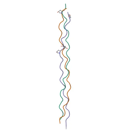
Polyclonal Antibodies
Anti-Collagen I/COL1A2 Polyclonal Antibody for WB, IF/ICC, ELISA - P08123
Item Number : CM0021019
Price varies based on specs and customizations
- Application
- WB, IF/ICC, ELISA
- Cross Reactivity
- Human, Mouse, Rat
- Protein Weight
- 129kDa

Shipping:
Contact us to get shipping details Enjoy On-time Dispatch Guarantee.
Why Choose Us
Easy ordering process, quality products, and dedicated support for your business success.
Core Product Specifications and Parameters
| Parameter | Value |
|---|---|
| Product Name | Collagen I/COL1A2 Rabbit pAb |
| Remarks/Alias | OI4; EDSCV; EDSARTH2; Collagen I/COL1A2 |
| Species | Human |
| GeneID | 1278 (Human) |
| Immunogen | Synthetic peptide corresponding to a sequence within amino acids 1161-1260 of human Collagen I/COL1A2 (NP_000080.2) |
| Source | Rabbit |
| Category | Polyclonal Antibodies |
| Application | WB, IF/ICC, ELISA |
| Cross Reactivity | Human, Mouse, Rat |
| SWISS | P08123 |
| Protein Weight | 129kDa |
| Shipping | Ice bag |
Biological Background: Collagen alpha-2(I) Chain Function and Localization
- Collagen alpha-2(I) chain is a subunit of type I collagen, the most abundant fibrillar collagen in the human body, providing tensile strength to connective tissues.
- Together with two alpha-1(I) chains, it forms the heterotrimeric type I procollagen molecule, which undergoes proteolytic processing to assemble into mature collagen fibrils.
- Type I collagen is a major structural component of the extracellular matrix in skin, bone, tendons, ligaments, and other connective tissues, where it organizes into fibrils that are mineralized with calcium hydroxyapatite in bones.
- The protein is synthesized as a preproprotein with a signal peptide, and the mature chain is secreted into the extracellular space, contributing to the supramolecular assembly of collagen fibers.
- Tissue-specific expression: highly expressed in tendon, ligament, and bone, with fibril organization critical for biomechanical properties; mutations in COL1A2 cause osteogenesis imperfecta and Ehlers-Danlos syndrome.
- Post-translational modifications include hydroxylation of proline and lysine residues, glycosylation, and formation of disulfide bonds, which are essential for triple helix stability and fibril cross-linking.
- The protein contains a large triple-helical domain with characteristic Gly-X-Y repeats, where X is often proline and Y is often hydroxyproline, and plays a role in cell adhesion via integrin-binding sites.
Experimental Guidance and Technical Tips
- The immunogen is a synthetic peptide from the C-terminal region (aa 1161-1260) of human COL1A2, which may recognize both the pro-form and mature collagen; validate in your sample system.
- For Western blot, use reducing SDS-PAGE conditions to dissociate collagen trimers; note that the theoretical molecular weight of the pro-alpha-2 chain is ~129 kDa, but mature processed forms may appear smaller.
- When performing immunofluorescence (IF/ICC), consider that collagen is secreted; if staining intracellular procollagen, include permeabilization steps and co-localize with ER/Golgi markers.
- ELISA applications may be used to detect soluble collagen fragments or secreted collagen in cell culture supernatants; optimize coating and detection conditions for your matrix.
- The antibody cross-reacts with mouse and rat, supporting translational studies in animal models of fibrosis, bone disease, and wound healing.
CamelBio: Your One-Stop Sourcing Bridge
CamelBio empowers diagnostic and research communities with streamlined access to premium IVD raw materials. From validated antibody pairs and optimized polyclonal antibodies like this anti-Collagen I/COL1A2 pAb to bulk ancillary reagents and rare-target sourcing, we bridge your project from concept to clinic. Whether your focus is extracellular matrix biology, bone metabolism, or connective tissue disorders, our one-stop supply services help accelerate your assay development.
Product Datasheet
Anti-Collagen I/COL1A2 Polyclonal Antibody for WB, IF/ICC, ELISA - P08123
REQUEST A QUOTE
Our professional team will reply to you within one business day. Please feel free to contact us!