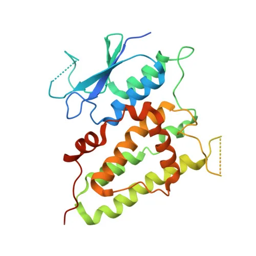
Polyclonal Antibodies
Anti-CLIC4 Rabbit Polyclonal Antibody for WB, IHC-P, IF/ICC, ELISA - Q9Y696
Item Number : CM0028391
Price varies based on specs and customizations
- Application
- WB, IHC-P, IF/ICC, ELISA
- Cross Reactivity
- Human, Mouse, Rat
- Protein Weight
- 29kDa

Shipping:
Contact us to get shipping details Enjoy On-time Dispatch Guarantee.
Why Choose Us
Easy ordering process, quality products, and dedicated support for your business success.
Core Product Specifications and Parameters
| Parameter | Value |
|---|---|
| Product Name | CLIC4 Rabbit pAb |
| Remarks/Alias | H1; huH1; p64H1; CLIC4L; MTCLIC; CLIC4 |
| Species | Human |
| GeneID (Human) | 25932 |
| GeneID | 25932 |
| Immunogen | Recombinant fusion protein containing a sequence corresponding to amino acids 1-253 of human CLIC4 (NP_039234.1) |
| Source | Rabbit |
| Category | Polyclonal Antibodies |
| Application | WB, IHC-P, IF/ICC, ELISA |
| Cross Reactivity | Human, Mouse, Rat |
| SWISS | Q9Y696 |
| Protein Weight | 29kDa |
| Shipping | Ice bag |
Biological Background: CLIC4 Function and Localization
- CLIC4 (Chloride intracellular channel protein 4), also known as p64H1, H1, huH1, MTCLIC, and CLIC4L, is a 253-amino acid protein belonging to the chloride intracellular channel (CLIC) family.
- In its soluble form, CLIC4 functions as a glutaredoxin-like oxidoreductase, catalyzing thiol-disulfide exchange reactions using reduced glutathione as an electron donor (PubMed:25581026, PubMed:37759794). Related references: PMID:25581026 PMID:37759794
- Upon membrane insertion, which may be redox-regulated, CLIC4 forms voltage-dependent multi-ion conductive channels selective for chloride and other anions (PubMed:16176272). Related references: PMID:16176272
- Beyond ion transport, CLIC4 has been implicated in angiogenesis, endothelial cell proliferation, and endothelial morphogenesis (tubulogenesis), potentially through membrane trafficking and cell-surface receptor modulation (e.g., HRH3).
- Subcellular localization is diverse: CLIC4 is found in the cytoplasm, nucleus, cytoskeleton (centrosome), cytoplasmic vesicle membranes, cell membrane, mitochondria, cell junctions, and endoplasmic reticulum membrane, reflecting its dynamic roles.
- Tissue expression is broad: detected at protein level in epithelial cells of colon, esophagus, and kidney; mRNA is prominent in heart, kidney, placenta, and skeletal muscle.
- The protein undergoes post-translational modifications including acetylation and phosphorylation, and its activity is regulated by redox state, as highlighted by UniProt keywords: Acetylation, Phosphoprotein, Redox-active.
Experimental Guidance and Technical Tips
- The immunogen covers residues 1–253 (full-length human CLIC4), so the resulting polyclonal antibody may recognize the N-terminal domain; validate detection of recombinant and endogenous CLIC4 in your model system.
- For Western blotting, a band around 29 kDa is expected under reducing conditions; additional bands may arise due to post-translational modifications or protein complexes.
- For immunohistochemistry (IHC-P) and immunofluorescence (IF/ICC), fixative and antigen retrieval optimization are recommended; citrate or EDTA-based protocols are typical starting points.
- Cross-reactivity with mouse and rat CLIC4 is reported; sequence alignment of the immunogen region can help assess suitability for other species.
- Consider using the antibody in ELISA for quantitative detection; titration of antibody and antigen concentrations is advised for optimal signal-to-noise ratio.
CamelBio: Your One-Stop Sourcing Bridge
CamelBio is your one-stop sourcing partner for high-quality IVD raw materials, including validated polyclonal antibodies like anti-CLIC4 for ion channel and oxidoreductase research. We supply optimized antibody reagents, bulk ancillary materials, and can help source rare-target raw materials to streamline your diagnostic development pipeline.
Product Datasheet
Anti-CLIC4 Rabbit Polyclonal Antibody for WB, IHC-P, IF/ICC, ELISA - Q9Y696
REQUEST A QUOTE
Our professional team will reply to you within one business day. Please feel free to contact us!