Polyclonal Antibodies
Anti-CFI Polyclonal Antibody for WB and ELISA - P05156
Item Number : CM0026615
Price varies based on specs and customizations
- Application
- WB, ELISA
- Cross Reactivity
- Human, Mouse
- Protein Weight
- 66kDa

Shipping:
Contact us to get shipping details Enjoy On-time Dispatch Guarantee.
Why Choose Us
Easy ordering process, quality products, and dedicated support for your business success.
Core Product Specifications and Parameters
| Parameter | Value |
|---|---|
| Product Name | CFI Rabbit pAb |
| Remarks/Alias | FI; IF; KAF; AHUS3; ARMD13; C3BINA; C3b-INA; CFI |
| Species | Human |
| GeneID (Human) | 3426 |
| GeneID | 3426 |
| Immunogen | Recombinant fusion protein containing a sequence corresponding to amino acids 19-300 of human CFI (NP_000195.2). |
| Source | Rabbit |
| Category | Polyclonal Antibodies |
| Application | WB, ELISA |
| Cross Reactivity | Human, Mouse |
| SWISS | P05156 |
| Protein Weight | 66kDa |
| Shipping | Ice bag |
Biological Background: CFI Function and Localization
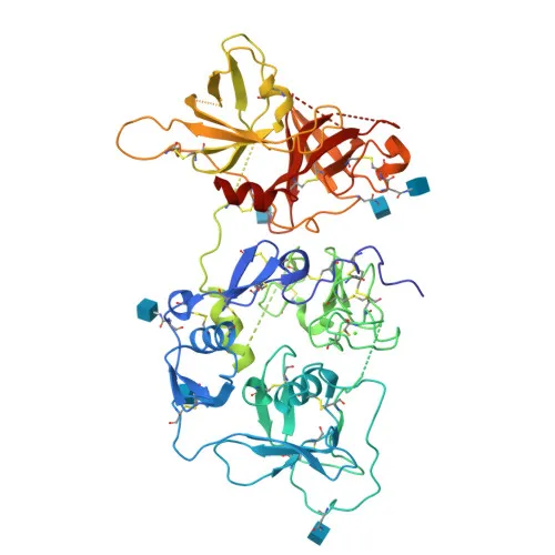
- CFI is a trypsin-like serine protease that negatively regulates all complement pathways by cleaving the alpha-chains of C3b and C4b, thereby inactivating these key opsonins.
Related references: PMID:17320177 PMID:7360115 - It requires cofactors for activity, including factor H and C4BP in the fluid phase, and membrane cofactor protein (CD46) and complement receptor 1 (CR1) on cell surfaces.
Related references: PMID:12055245 PMID:2141838 PMID:9605165 - On healthy host cells, CFI-mediated degradation of deposited C3b prevents autologous complement activation, while on apoptotic cells or microbes lacking protective cofactors, C3b remains active to promote opsonization and clearance.
Related references: PMID:28671664 - CFI is primarily expressed in the liver by hepatocytes, but is also found in monocytes, fibroblasts, and keratinocytes.
Related references: PMID:6327681 PMID:17320177 PMID:6444659 - The protein is secreted into the extracellular space and circulates in the blood, exerting its regulatory function in the fluid phase and on cell surfaces.
- CFI contains disulfide bonds, glycosylation sites, and calcium-binding motifs; it is classified under serine proteases and is involved in innate immunity. Mutations are associated with age-related macular degeneration and hemolytic uremic syndrome.
- The protein has a predicted molecular mass of ~66 kDa and undergoes N-linked glycosylation.
Experimental Guidance and Technical Tips
- The immunogen (aa 19-300) spans the N-terminal portion of human CFI, likely recognizing both pro‑CFI and the mature heavy chain; consider this when interpreting band patterns.
- For Western blot, recommended starting dilution is typically 1:500–1:2000; an expected band near 66 kDa should be observed under reducing conditions, with possible additional bands from glycosylation or processing.
- For ELISA, test conditions require optimization; the antibody can be used for antigen capture or detection, but validate pairing with a complementary antibody if using a sandwich format.
- When analyzing mouse samples, verify reactivity; while cross-reactivity is listed, signal intensity may differ from human CFI.
- Always include appropriate positive and negative controls, such as lysates from CFI-expressing cells (e.g., hepatocyte-derived cell lines) and CFI-depleted serum.
CamelBio: Your One-Stop Sourcing Bridge
CamelBio provides diagnostic manufacturers, labs, and research institutes with one-stop access to IVD raw materials, technical services, and consulting—covering every stage from concept to clinic. For complement research and assay development, CamelBio can source validated antibody pairs, optimized polyclonal antibodies, bulk ancillary reagents, and rare-target raw materials to streamline your workflow.
REQUEST A QUOTE
Our professional team will reply to you within one business day. Please feel free to contact us!