Polyclonal Antibodies
Anti-CDKN1B/p27KIP1 Polyclonal Antibody for WB, IF/ICC, IHC-P, ELISA - P46527
Item Number : CM0017717
Price varies based on specs and customizations
- Application
- WB, IF/ICC, IHC-P, ELISA
- Cross Reactivity
- Human, Mouse, Rat
- Protein Weight
- 22kDa

Shipping:
Contact us to get shipping details Enjoy On-time Dispatch Guarantee.
Why Choose Us
Easy ordering process, quality products, and dedicated support for your business success.
Core Product Specifications and Parameters
| Parameter | Value |
|---|---|
| Product Name | CDKN1B/p27KIP1 Rabbit pAb |
| Remarks/Alias | KIP1; MEN4; CDKN4; MEN1B; P27KIP1; CDKN1B/p27KIP1 |
| Species | Human |
| GeneID (Human) | 1027 |
| GeneID | 1027 |
| Immunogen | Recombinant protein|Recombinant fusion protein containing a sequence corresponding to amino acids 1-198 of human CDKN1B/p27KIP1 (NP_004055.1). |
| Source | Rabbit |
| Category | Polyclonal Antibodies |
| Application | WB, IF/ICC, IHC-P, ELISA |
| Cross Reactivity | Human, Mouse, Rat |
| SWISS | P46527 |
| Protein Weight | 22kDa |
| Shipping | Ice bag |
Biological Background: CDKN1B/p27KIP1 Function and Localization
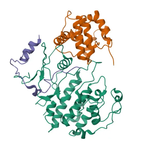
- CDKN1B encodes cyclin-dependent kinase inhibitor 1B, also known as p27Kip1, a key regulator of cell cycle progression, primarily mediating G1 arrest.
- It inhibits cyclin E-CDK2 and cyclin A-CDK2 complexes, thereby blocking cell cycle progression, but has little inhibitory effect on CDK2 bound to SPDYA. Related references: PMID:28666995
- p27Kip1 also interacts with cyclin D-CDK4 complexes, modulating their assembly, stability, and activation; its effect can be either inhibitory or activating depending on phosphorylation state and stoichiometry.
- Subcellular localization includes nucleus, cytoplasm, and endosomes, reflecting dynamic regulation during the cell cycle.
- Post-translational modifications, particularly phosphorylation, control p27Kip1 activity and subcellular distribution (Keywords: Phosphoprotein).
- It is expressed in all tissues tested, with highest levels in skeletal muscle and lowest in liver and kidney; protein expression confirmed in kidney. Related references: PMID:15509543 PMID:8033212
- Functionally, CDKN1B acts as a tumor suppressor, and its dysregulation is implicated in multiple cancers (Keywords: Tumor suppressor).
- The protein undergoes ubiquitin-like (Ubl) conjugation and is identified by proteomics, indicating post-translational control mechanisms.
Experimental Guidance and Technical Tips
- The immunogen spans the full-length human p27Kip1 (amino acids 1-198), encompassing known functional domains; the antibody may detect both phosphorylated and unphosphorylated forms — consider phosphatase treatment in relevant assays.
- For Western blotting, a band at approximately 22 kDa is expected; optimize antibody concentration and blocking conditions for your specific sample.
- In immunostaining (IF/ICC, IHC-P), note that p27Kip1 can localize to the nucleus, cytoplasm, or endosomes depending on cell cycle stage and modifications; validate subcellular distribution in your model system.
- As the antibody cross-reacts with mouse and rat, it is suitable for rodent studies; however, verify species-specific reactivity and background in your experimental setup.
- For ELISA, ensure appropriate coating and detection conditions; this antibody can be used for direct or indirect detection applications.
CamelBio: Your One-Stop Sourcing Bridge
CamelBio serves as a comprehensive sourcing bridge for IVD raw materials, connecting diagnostic manufacturers and research labs with high-quality reagents. For cell cycle and oncology research, we offer validated antibody pairs, optimized polyclonals like this anti-CDKN1B/p27KIP1 antibody, and bulk ancillary reagents to support assay development. If you're exploring rare or challenging targets, our sourcing network can help secure customized immunoreagents for your pipeline.
Product Datasheet
Anti-CDKN1B/p27KIP1 Polyclonal Antibody for WB, IF/ICC, IHC-P, ELISA - P46527
REQUEST A QUOTE
Our professional team will reply to you within one business day. Please feel free to contact us!