Polyclonal Antibodies
Anti-CDH3 Rabbit Polyclonal Antibody for WB, IHC-P, IF/ICC, ELISA - P22223
Item Number : CM0017908
Price varies based on specs and customizations
- Application
- WB, IHC-P, IF/ICC, ELISA
- Cross Reactivity
- Human, Mouse, Rat
- Protein Weight
- 91kDa

Shipping:
Contact us to get shipping details Enjoy On-time Dispatch Guarantee.
Why Choose Us
Easy ordering process, quality products, and dedicated support for your business success.
Core Product Specifications and Parameters
| Parameter | Value |
|---|---|
| Product Name | CDH3 Rabbit pAb |
| Remarks/Alias | CDHP; HJMD; PCAD; CDH3 |
| Species | Human |
| GeneID (human) | 1001 |
| GeneID | 1001 |
| Immunogen | Recombinant fusion protein containing a sequence corresponding to amino acids 24-350 of human CDH3 (NP_001784.2) |
| Source | Rabbit |
| Category | Polyclonal Antibodies |
| Application | WB, IHC-P, IF/ICC, ELISA |
| Cross Reactivity | Human, Mouse, Rat |
| SWISS | P22223 |
| Protein Weight | 91kDa |
| Shipping | Ice bag |
Biological Background: Cadherin-3 Function and Localization
- Cadherin-3, also known as placental cadherin (P-cadherin), is a classical cadherin encoded by the CDH3 gene (gene aliases: CDHP). It is classified as a calcium-dependent cell adhesion protein.
- As a member of the cadherin family, CDH3 mediates cell–cell adhesion through homophilic interactions, contributing to the sorting of heterogeneous cell types and maintenance of tissue architecture.
- The protein is localized to the cell membrane, consistent with its role as a transmembrane adhesion receptor.
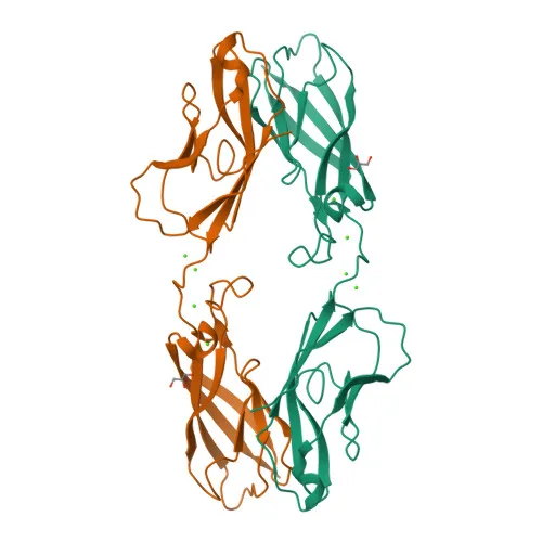
- Key structural features include cadherin repeat domains, a signal peptide, and a cleavage site for paired basic residues, suggesting post-translational processing. It is also reported to be glycosylated.
- Tissue expression is observed in some normal epithelial tissues and certain carcinoma cell lines, indicating potential roles in both normal epithelial biology and tumor progression.
- The cadherin-3 protein has a molecular weight of approximately 91–92 kDa (predicted 91,418 Da), with possible proteolytic fragments.
Experimental Guidance and Technical Tips
- The immunogen (amino acids 24–350) spans the N-terminal extracellular cadherin repeat region; this design may detect both full-length CDH3 and cleaved extracellular domains.
- For Western blot, the predicted molecular weight is ~91 kDa; under reducing conditions, use appropriate gel electrophoresis and transfer protocols.
- In IHC-P, antigen retrieval methods may be required; optimization for your specific tissue and fixation conditions is recommended.
- For IF/ICC, include a permeabilization step (e.g., Triton X-100) to access membrane-localized CDH3; co-staining with a membrane marker may confirm specificity.
- ELISA sensitivity depends on coating conditions and detection reagents; consider checkerboard titration for optimal signal-to-noise ratio.
- Although cross-reactivity with mouse and rat is reported, validation in the intended sample system is essential to confirm species-specific performance.
CamelBio: Your One-Stop Sourcing Bridge
CamelBio provides diagnostic manufacturers, contract research organizations, and academic laboratories with streamlined access to validated IVD raw materials—including polyclonal and monoclonal antibodies, recombinant proteins, and ancillary reagents. This anti-CDH3 antibody supports your cell adhesion and epithelial biology research or assay development, with flexible supply options from research-scale to bulk orders. To accelerate your diagnostic pipeline from concept to clinic, CamelBio also offers technical consulting and rare-target sourcing services.
Product Datasheet
Anti-CDH3 Rabbit Polyclonal Antibody for WB, IHC-P, IF/ICC, ELISA - P22223
REQUEST A QUOTE
Our professional team will reply to you within one business day. Please feel free to contact us!