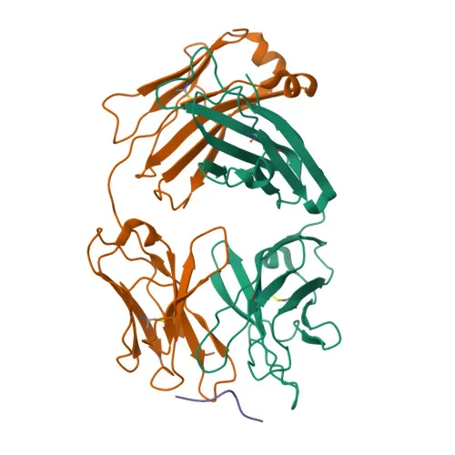
Monoclonal Antibodies
Anti-CD99 Rabbit Monoclonal Antibody for WB, IF/ICC, IHC-P, FC, ELISA - P14209
Item Number : CM0010785
Price varies based on specs and customizations
- Application
- WB, IF/ICC, IHC-P, FC, ELISA
- Cross Reactivity
- Human
- Protein Weight
- 16-19kDa

Shipping:
Contact us to get shipping details Enjoy On-time Dispatch Guarantee.
Why Choose Us
Easy ordering process, quality products, and dedicated support for your business success.
Core Product Specifications and Parameters
| Parameter | Value |
|---|---|
| Product Name | CD99 Rabbit mAb |
| Remarks/Alias | MIC2; HBA71; MIC2X; MIC2Y; MSK5X |
| Species | Human |
| GeneID (Human) | 4267 |
| GeneID | 4267 |
| Immunogen | Synthetic peptide - A synthetic peptide corresponding to a sequence within amino acids 24-123 of human CD99 (NP_002405.1). |
| Source | Rabbit |
| Category | Monoclonal Antibodies |
| Application | WB, IF/ICC, IHC-P, FC, ELISA |
| Cross Reactivity | Human |
| SWISS | P14209 |
| Protein Weight | 16-19kDa |
| Shipping | Ice bag |
Biological Background: CD99 Function and Localization
- CD99, also known as MIC2, HBA71, MIC2X, MIC2Y, MSK5X, is a cell surface glycoprotein encoded by the CD99 gene (also known as MIC2, MIC2X, MIC2Y). It was originally identified as the E2 antigen on T-cells.
- The protein is involved in T-cell adhesion processes and spontaneous rosette formation with erythrocytes, playing a role in a late step of leukocyte extravasation helping leukocytes to overcome the endothelial basement membrane, acting independently of PECAM1.
- CD99 undergoes alternative splicing, producing multiple isoforms that may have distinct functions.
- Subcellular localization: CD99 is a transmembrane protein predominantly found on the plasma membrane.
- Post-translational modifications include glycosylation and phosphorylation, which may regulate its adhesive functions.
- The core protein has a predicted molecular weight of approximately 18.8 kDa, but due to glycosylation, it migrates at 16-19 kDa in SDS-PAGE.
- CD99 is expressed in a wide range of hematopoietic cells and is used as a diagnostic marker for Ewing sarcoma and primitive neuroectodermal tumors.
Experimental Guidance and Technical Tips
- Given that the immunogen corresponds to the N-terminal extracellular domain (amino acids 24-123), the antibody is likely to recognize the native, non-permeabilized form of CD99, making it suitable for surface staining in flow cytometry and immunofluorescence.
- For Western blotting, expect a band around 16-19 kDa; using reducing conditions may help resolve the glycosylated forms. Validate specificity with positive and negative cell lines or siRNA knockdown.
- In IHC-P, antigen retrieval (e.g., heat-induced epitope retrieval in citrate buffer) may improve detection; titration is recommended.
- For IF/ICC, both fixed and live-cell staining can be tested; if using fixation, gentle permeabilization may be optional since the epitope is extracellular.
- Always include appropriate isotype controls and confirm cross-reactivity only to human, as per product specification.
CamelBio: Your One-Stop Sourcing Bridge
CamelBio is a dedicated IVD raw material partner, offering end-to-end support from concept to clinic for diagnostic manufacturers and research laboratories. This CD99 rabbit monoclonal antibody is part of our expanding immunology and oncology portfolio, designed to accelerate assay development. Beyond individual antibodies, we provide validated antibody pairs, bulk ancillary reagents, and customized sourcing for rare targets, ensuring you have reliable materials for your workflow.
Product Datasheet
Anti-CD99 Rabbit Monoclonal Antibody for WB, IF/ICC, IHC-P, FC, ELISA - P14209
REQUEST A QUOTE
Our professional team will reply to you within one business day. Please feel free to contact us!