Polyclonal Antibodies
Anti-CD99 Polyclonal Antibody for WB, ELISA - P14209
Item Number : CM0023366
Price varies based on specs and customizations
- Application
- WB, ELISA
- Cross Reactivity
- Human
- Protein Weight
- 19kDa

Shipping:
Contact us to get shipping details Enjoy On-time Dispatch Guarantee.
Why Choose Us
Easy ordering process, quality products, and dedicated support for your business success.
Core Product Specifications and Parameters
| Parameter | Value |
|---|---|
| Product Name | CD99 Rabbit pAb |
| Remarks/Alias | MIC2; HBA71; MIC2X; MIC2Y; MSK5X; CD99 |
| Species | Human |
| GeneID (human) | 4267 |
| GeneID | 4267 |
| Immunogen | Synthetic peptide corresponding to a sequence within amino acids 23-124 of human CD99 (NP_002405.1) |
| Source | Rabbit |
| Category | Polyclonal Antibodies |
| Application | WB, ELISA |
| Cross Reactivity | Human |
| SWISS | P14209 |
| Protein Weight | 19kDa |
| Shipping | Ice bag |
Biological Background: CD99 Function and Localization
- CD99, also known as MIC2 or T-cell surface glycoprotein E2, is a heavily glycosylated transmembrane protein that belongs to the CD99 family. It undergoes alternative splicing, generating multiple isoforms.
- The protein is primarily expressed on the surface of T-cells, where it mediates cell-cell adhesion. It is involved in spontaneous rosette formation between T-cells and erythrocytes, a classic immunological phenomenon.
- CD99 plays a critical role in leukocyte extravasation, specifically in the step where leukocytes transmigrate through the endothelial basement membrane. It functions at the same site as but independently of PECAM-1.
- The protein is localized to the plasma membrane as a type I transmembrane protein, with an extracellular domain mediating adhesive interactions and a short cytoplasmic tail implicated in signaling.
- CD99 is subject to post-translational modifications including phosphorylation and extensive glycosylation, which modulate its function and molecular weight. The mature glycoprotein runs at approximately 32 kDa on SDS-PAGE, though the core protein weight is around 19 kDa.
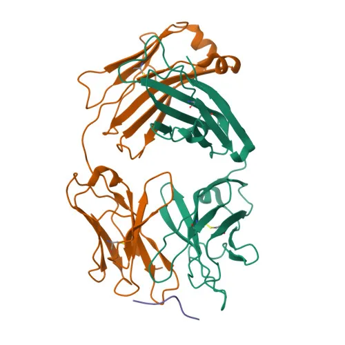
- Structural studies (3D-structure) have provided insights into the molecular basis of CD99-mediated adhesion, and direct protein sequencing has confirmed its identity and processing.
- Although best characterized in T-cells, CD99 is also expressed on other leukocytes and endothelial cells, contributing to immune surveillance and inflammatory responses.
Experimental Guidance and Technical Tips
- The immunogen for this polyclonal antibody corresponds to the N-terminal extracellular region (aa 23–124), which includes part of the adhesion domain. For WB, this antibody may detect bands corresponding to differentially glycosylated isoforms; consider using a deglycosylation step to confirm the core protein size.
- In ELISA applications, the antibody can be used to capture or detect native CD99 in cell lysates or purified protein preparations. Optimization of coating and blocking conditions is recommended to minimize non-specific binding.
- For tissue or cell staining, validate the antibody in the relevant human sample system (e.g., Jurkat T-cells, peripheral blood mononuclear cells) and include appropriate controls such as isotype-matched IgG or peptide-blocked samples to confirm specificity.
CamelBio: Your One-Stop Sourcing Bridge
CamelBio supplies comprehensive IVD raw materials and technical support for diagnostic developers and research laboratories. For projects targeting immune cell adhesion molecules like CD99, we offer validated antibody pairs, optimized monoclonal and polyclonal antibodies, and bulk ancillary reagents. Our platform streamlines procurement from early-stage research to clinical assay development, ensuring reliable access to rare-target raw materials and customized sourcing solutions.
REQUEST A QUOTE
Our professional team will reply to you within one business day. Please feel free to contact us!