Monoclonal Antibodies
Anti-CD96 Rabbit Monoclonal Antibody for WB, FC, ELISA - P40200
Item Number : CM0007589
Price varies based on specs and customizations
- Application
- WB, FC, ELISA
- Cross Reactivity
- Human
- Protein Weight
- 65kDa

Shipping:
Contact us to get shipping details Enjoy On-time Dispatch Guarantee.
Why Choose Us
Easy ordering process, quality products, and dedicated support for your business success.
Core Product Specifications and Parameters
| Parameter | Value |
|---|---|
| Product Name | CD96 Rabbit mAb |
| Remarks/Alias | Cell surface antigen CD96; CD96 |
| Species | Human |
| GeneID (Human) | 10225 |
| GeneID | 10225 |
| Immunogen | Recombinant fusion protein containing a sequence corresponding to amino acids 25-519 of human CD96 (NP_937839.1) |
| Source | Rabbit |
| Category | Monoclonal Antibodies |
| Application | WB, FC, ELISA |
| Cross Reactivity | Human |
| SWISS | P40200 |
| Protein Weight | 65kDa |
| Shipping | Ice bag |
Biological Background: CD96 Function and Localization
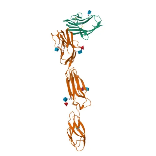
- CD96, also known as T-cell surface protein tactile or cell surface antigen CD96, is a transmembrane glycoprotein belonging to the immunoglobulin superfamily involved in cell adhesion and immune regulation.
- Mediates adhesive interactions of activated T cells and NK cells during late-stage immune responses, promoting NK cell-target adhesion by binding to poliovirus receptor (PVR/CD155) on target cells.
- Functions after lymphocyte extravasation, facilitating engagement with diseased cells and movement within inflammatory sites, complementing integrin- and selectin-mediated adhesion.
- Encoded by the CD96 gene (GeneID: 10225), with alternative splicing producing multiple isoforms; the canonical isoform has a predicted molecular weight of approximately 65.6 kDa (UniProt: P40200).
- Predominantly expressed on the plasma membrane; tissue expression is mainly on normal T-cell lines, clones, and some transformed T-cells, with very low levels on activated B cells.
- Contains an extracellular region with immunoglobulin-like domains, a single transmembrane helix, and a cytoplasmic tail; features disulfide bond formation and glycosylation.
- Associated with disease variants, including chromosomal rearrangements linked to craniosynostosis, and identified in proteomics screens as a reference proteome component.
Experimental Guidance and Technical Tips
- The immunogen corresponds to the extracellular domain (amino acids 25-519), suggesting reactivity against the native surface protein; consider using live cell staining in flow cytometry to preserve epitope conformation.
- Predicted molecular weight of CD96 is 65 kDa; due to glycosylation, the observed band in Western blot may appear slightly higher; include a positive control lysate from T-cell lines.
- For ELISA, coating with recombinant PVR may enable detection of functional antibody binding, but direct antigen coating with CD96 protein is standard.
- Specificity should be confirmed using CD96-overexpressing cells or siRNA knockdown in relevant samples.
- This antibody is reported for human CD96; cross-reactivity with other species has not been determined; perform a sequence alignment check before testing on non-human samples.
CamelBio: Your One-Stop Sourcing Bridge
CamelBio provides diagnostic manufacturers and research laboratories with one-stop access to high-quality IVD raw materials. For CD96-related immunoassay development, we offer validated monoclonal antibodies like this CD96 Rabbit mAb, along with potential antibody pairs, recombinant proteins, and ancillary reagents to accelerate your project from concept to clinic. Our sourcing network can also accommodate rare target requests to fill gaps in your development pipeline.
REQUEST A QUOTE
Our professional team will reply to you within one business day. Please feel free to contact us!