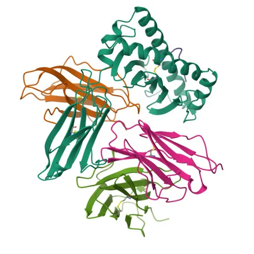
Monoclonal Antibodies
Anti-CD8a Monoclonal Antibody for Flow Cytometry - P01732
Item Number : CM0009129
Price varies based on specs and customizations
- Application
- Flow Cytometry (FC)
- Cross Reactivity
- Human, Cynomolgus
- Protein Weight
- 21kDa/25kDa/30kDa

Shipping:
Contact us to get shipping details Enjoy On-time Dispatch Guarantee.
Why Choose Us
Easy ordering process, quality products, and dedicated support for your business success.
Core Product Specifications and Parameters
| Parameter | Value |
|---|---|
| Product Name | Rabbit anti-Human/Monkey CD8a mAb |
| Remarks/Alias | CD8; p32; Leu2; CD8alpha |
| Species | Human |
| Gene ID (Human) | 925 |
| Gene ID | 925 |
| Immunogen | Recombinant fusion protein containing a sequence corresponding to amino acids 22-182 of human CD8A (NP_001759.3) |
| Source | Rabbit |
| Category | Monoclonal Antibodies |
| Application | Flow Cytometry (FC) |
| Cross Reactivity | Human, Cynomolgus |
| SWISS | P01732 |
| Protein Weight | 21kDa/25kDa/30kDa |
| Shipping | Ice bag |
Biological Background: CD8a Function and Localization
- CD8a is a T-cell surface glycoprotein that serves as a coreceptor for MHC class I molecules, enabling cytotoxic T-lymphocytes to recognize and eliminate infected or tumor cells.
- It exists as either a heterodimer with CD8B or a homodimer, and is expressed on thymocytes, cytotoxic T-cells, NK cells, memory T-cells, and intraepithelial lymphocytes.
- CD8a simultaneously engages the T-cell receptor (TCR) and MHC class I proteins, recruiting the LCK kinase to phosphorylate signaling substrates for T-cell activation, cytokine production, and adhesion.
- In NK-cells, CD8A homodimers support survival and serial killing of target cells, while in activated lymphocytes they promote differentiation into memory CD8+ T-cells.
- The protein is a single-pass type I membrane protein, but a secreted isoform exists; alternative splicing generates multiple transcripts.
- Post-translational modifications include disulfide bond formation, N-glycosylation, and palmitoylation, which anchor it to lipid rafts.
- Tissue specificity: expressed on a subset of immune cells, and upregulated on plasmacytoid dendritic cells upon herpes simplex virus-1 stimulation.
Experimental Guidance and Technical Tips
- The immunogen corresponds to the extracellular domain (aa 22–182) of human CD8A, making this antibody suitable for detecting surface CD8a by flow cytometry.
- For flow cytometry, use standard surface staining protocols with appropriately titrated antibody; consider using live/dead discrimination and appropriate isotype controls.
- Validate reactivity in relevant cell systems such as human PBMCs or cynomolgus monkey lymphocytes, as cross-reactivity is indicated.
- Depending on experimental design, fixation/permeabilization steps may be optimized if intracellular staining is required.
- For multiplex panels, check fluorophore compatibility and potential steric hindrance with other markers in the CD8 co-receptor complex.
CamelBio: Your One-Stop Sourcing Bridge
CamelBio supports diagnostic manufacturers and research laboratories by providing one-stop access to IVD raw materials, including validated antibody pairs, optimized monoclonal/polyclonal antibodies, and bulk ancillary reagents. We specialize in immunology targets like CD8a for T-cell profiling, offering reliable supply chains from concept to clinic. For rare or custom targets, our sourcing services bridge the gap between discovery and commercial-scale production, ensuring you get the critical reagents you need.
REQUEST A QUOTE
Our professional team will reply to you within one business day. Please feel free to contact us!