Monoclonal Antibodies
Anti-CD5 Rabbit Monoclonal Antibody for WB, IHC-P, IF/ICC, ELISA - P06127
Item Number : CM0011235
Price varies based on specs and customizations
- Application
- WB, IHC-P, IF/ICC, ELISA
- Cross Reactivity
- Human
- Protein Weight
- 55kDa

Shipping:
Contact us to get shipping details Enjoy On-time Dispatch Guarantee.
Why Choose Us
Easy ordering process, quality products, and dedicated support for your business success.
Core Product Specifications and Parameters
| Parameter | Value |
|---|---|
| Product Name | CD5 Rabbit mAb |
| Remarks/Alias | T1; LEU1; CD5 |
| Species | Human |
| GeneID (Human) | 921 |
| GeneID | 921 |
| Immunogen | Synthetic Peptide: A synthetic peptide corresponding to a sequence within amino acids 1-100 of human CD5 (P06127). |
| Source | Rabbit |
| Category | Monoclonal Antibodies |
| Application | WB, IHC-P, IF/ICC, ELISA |
| Cross Reactivity | Human |
| SWISS | P06127 |
| Protein Weight | 55kDa |
| Shipping | Ice bag |
Biological Background: CD5 Function and Localization
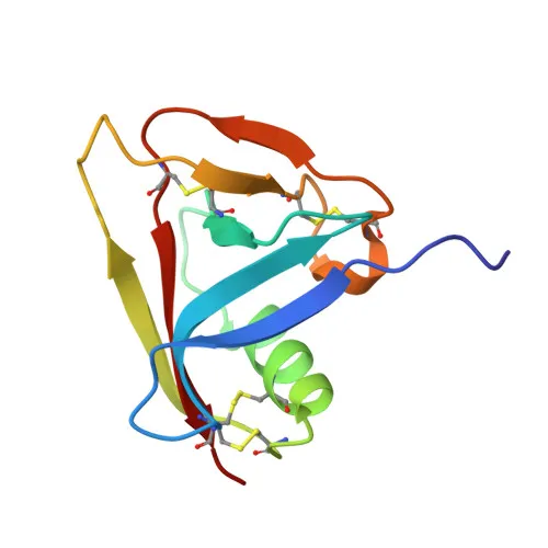
- CD5 (Cluster of Differentiation 5), also known as T1/Leu-1, is a type I transmembrane glycoprotein belonging to the scavenger receptor cysteine-rich (SRCR) superfamily.
- It is expressed on all T cells and a subset of B cells (B1a cells), functioning as a lymphoid-specific receptor that regulates TCR and BCR signaling, thymocyte selection, and immune tolerance.
- CD5 acts as a negative regulator of TCR signaling by associating with key signaling molecules such as LCK, CD3Z chain, PI3K, and CBL.
Related references: PMID:1384049, PMID:1385158 - Upon co-engagement with CD3, CD5 enhances CBL recruitment, leading to increased VAV1 phosphorylation and degradation, thereby modulating signal strength.
Related references: PMID:23376399 - In B cells, CD5 triggers a Ca(2+)-dependent ERK1/2 activation pathway via the non-selective Ca(2+) channel TRPC1, resulting in IL-10 production.
Related references: PMID:27499044 - CD5 interacts with ligands such as CD5L and CD72 on B cells, facilitating contact-mediated T-dependent B cell activation and maintaining homeostasis of regulatory T and B cells.
- Subcellularly, CD5 is localized to the cell membrane.
- The protein undergoes phosphorylation and glycosylation, contains disulfide bonds, a signal peptide, a transmembrane helix, and SRCR repeats. Its predicted molecular weight is approximately 55 kDa.
Experimental Guidance and Technical Tips
- The immunogen corresponds to amino acids 1–100 of human CD5, encompassing part of the extracellular region; this design may favor recognition of the native protein in various applications.
- For Western blot, consider using whole‑cell lysates from human T‑cell lines (e.g., Jurkat) or B1a cells; the expected band is ~55 kDa.
- In IHC‑P, optimize antigen retrieval and fixation using T‑cell‑rich tissues such as lymph node or spleen.
- For IF/ICC, test both paraformaldehyde and methanol fixation as well as permeabilization conditions to ensure proper membrane staining.
- Validate ELISA performance with the immunizing peptide or recombinant CD5 protein as a standard.
CamelBio: Your One-Stop Sourcing Bridge
CamelBio supports diagnostic manufacturers and research laboratories with one‑stop access to high‑quality IVD raw materials, including validated monoclonal antibodies like this anti‑CD5 clone. Backed by extensive supply capabilities—from optimized antibody pairs and bulk ancillary reagents to custom sourcing of rare targets—we help ensure your T‑cell and immunology projects move seamlessly from concept to clinic.
REQUEST A QUOTE
Our professional team will reply to you within one business day. Please feel free to contact us!