Monoclonal Antibodies
Anti-CD4 Monoclonal Antibody for Flow Cytometry - P01730
Item Number : CM0009694
Price varies based on specs and customizations
- Application
- FC
- Cross Reactivity
- Human, Cynomolgus

Shipping:
Contact us to get shipping details Enjoy On-time Dispatch Guarantee.
Why Choose Us
Easy ordering process, quality products, and dedicated support for your business success.
Core Product Specifications and Parameters
| Parameter | Value |
|---|---|
| Product Name | Rabbit anti-Human/Monkey CD4 mAb |
| Remarks/Alias | T4; IMD79; Leu-3; OKT4D; CD4mut |
| Species | Human |
| Gene ID (Human) | 920 |
| Gene ID | 920 |
| Immunogen | Recombinant protein of human CD4. |
| Source | Rabbit |
| Category | Monoclonal Antibodies |
| Application | FC |
| Cross Reactivity | Human, Cynomolgus |
| SWISS | P01730 |
| Protein Weight | 51kDa |
| Shipping | Ice bag |
Biological Background: CD4 Function and Localization
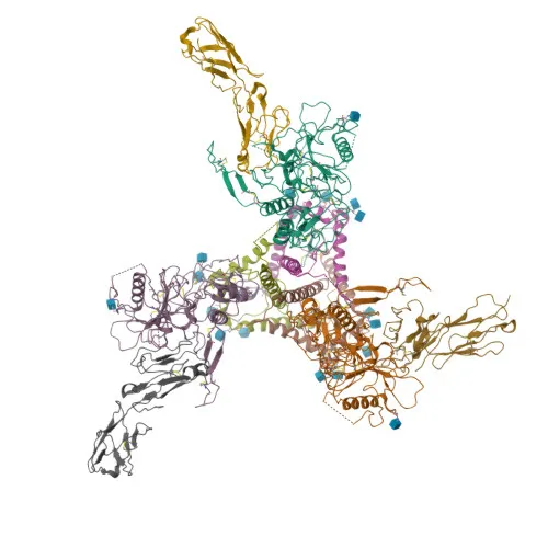
- CD4 is a transmembrane glycoprotein and member of the immunoglobulin superfamily, consisting of an extracellular region with four Ig-like domains (D1–D4), a single-pass transmembrane helix, and a short cytoplasmic tail. It is primarily expressed on the surface of T-helper cells, as well as on monocytes, macrophages, dendritic cells, and NK cells.
- In T-helper cells, CD4 functions as a coreceptor for MHC class II molecules. It binds to non-polymorphic regions of MHC class II on antigen-presenting cells, stabilizing the interaction between the T-cell receptor (TCR) and peptide-MHC complexes, which is essential for antigen recognition and T-cell activation.
- Upon TCR engagement, CD4 recruits the Src family kinase LCK via its cytoplasmic tail to the vicinity of the TCR-CD3 complex. LCK phosphorylates immunoreceptor tyrosine-based activation motifs (ITAMs) on CD3 chains, initiating intracellular signaling cascades that lead to T-cell proliferation, cytokine production, and differentiation.
- In addition to its role in adaptive immunity, CD4 acts as the primary receptor for human immunodeficiency virus type 1 (HIV-1) via interaction with the viral envelope glycoprotein gp120. It also serves as a receptor for human herpesvirus 7 (HHV-7). HIV-1 Vpu and Nef proteins downregulate CD4 surface expression to facilitate viral egress and immune evasion. Related references: PMID:12089508 PMID:16331979 PMID:2214026 PMID:17346169 PMID:7909607
- CD4 is post-translationally modified by N-linked glycosylation, disulfide bond formation, palmitoylation at a juxtamembrane cysteine, and phosphorylation by LCK. These modifications regulate protein folding, membrane localization, and signal transduction.
- The expression of CD4 is a defining characteristic of helper T cells and is commonly used as an immunophenotypic marker for T-cell subset identification. Its surface density can vary with cell activation and differentiation state.
- In macrophages and NK cells, CD4 engagement can trigger differentiation, cytokine expression, and cell migration through TCR/LCK-independent pathways, highlighting its functional versatility beyond T-cell signaling.
Experimental Guidance and Technical Tips
- This antibody is directly conjugated to , a bright and photostable far-red fluorophore, ready for use in flow cytometry. Titrate the antibody to determine the optimal staining concentration for your specific cell type and experimental conditions (typical range: 0.1–1 µg per 1×10^6 cells).
- Since CD4 surface expression is high on resting T-helper cells, include proper isotype controls and live/dead cell discrimination to ensure specific staining. For multicolor panels, combine with anti-CD3 and anti-CD8 to thoroughly dissect T-cell subsets.
- The antibody cross-reacts with both human and cynomolgus CD4, making it suitable for studies involving non-human primate models. Validate the antibody in your specific sample matrix and fixation/permeabilization protocol if intracellular staining is required.
- The immunogen is recombinant human CD4, likely corresponding to the extracellular domain. Therefore, the antibody may preferentially recognize native, non-denatured CD4; denaturing conditions (e.g., western blotting under reducing conditions) may require validation.
- For applications beyond flow cytometry, such as immunohistochemistry or immunoprecipitation, optimization and independent verification are recommended due to the lack of manufacturer data.
CamelBio: Your One-Stop Sourcing Bridge
CamelBio empowers diagnostic manufacturers and research laboratories by offering a comprehensive range of IVD raw materials, including validated antibodies, antigen pairs, and ancillary reagents. This CD4 antibody supports T-cell immunophenotyping and HIV-related research, and demonstrated reactivity in human and cynomolgus samples positions it for translational immunoassay development. Beyond individual reagents, CamelBio facilitates sourcing of rare-target raw materials and bulk custom orders, helping you streamline your entire workflow from assay design to clinical validation.
REQUEST A QUOTE
Our professional team will reply to you within one business day. Please feel free to contact us!