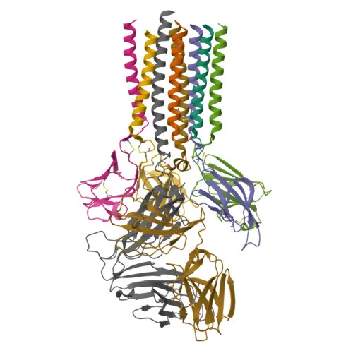
Polyclonal Antibodies
Anti-CD3G Rabbit Polyclonal Antibody for WB, ELISA - P09693
Item Number : CM0019561
Price varies based on specs and customizations
- Application
- WB, ELISA
- Cross Reactivity
- Human
- Protein Weight
- 20kDa

Shipping:
Contact us to get shipping details Enjoy On-time Dispatch Guarantee.
Why Choose Us
Easy ordering process, quality products, and dedicated support for your business success.
Core Product Specifications and Parameters
| Parameter | Value |
|---|---|
| Product Name | CD3G Rabbit pAb |
| Remarks/Alias | T3G; IMD17; CD3GAMMA; CD3-GAMMA; CD3G |
| Species | Human |
| Gene ID (Human) | 917 |
| Gene ID | 917 |
| Immunogen | Recombinant fusion protein containing a sequence corresponding to amino acids 23-116 of human CD3G (NP_000064.1) |
| Source | Rabbit |
| Category | Polyclonal Antibodies |
| Application | WB, ELISA |
| Cross Reactivity | Human |
| SWISS | P09693 |
| Protein Weight | 20kDa |
| Shipping | Ice bag |
Biological Background: CD3G Function and Localization
- CD3G encodes the T-cell surface glycoprotein CD3 gamma chain, also known as T3G, IMD17, or CD3-GAMMA; it belongs to the immunoglobulin superfamily and is a component of the TCR-CD3 complex.
- Upon TCR engagement by antigen-presenting cells, CD3G's cytoplasmic immunoreceptor tyrosine-based activation motifs (ITAMs) are phosphorylated by Src family kinases LCK and FYN, initiating downstream signaling pathways essential for T-cell activation. Related references: PMID:2470098
- CD3G contains a di-leucine (diL) receptor-sorting motif that regulates constitutive TCR cycling and surface expression, a mechanism critical for modulating T-cell responsiveness. Related references: PMID:8187769
- The protein is a single-pass type I membrane protein localized to the cell membrane; its extracellular region includes an immunoglobulin-like domain, and it undergoes N‑glycosylation and phosphorylation.
- Direct protein sequencing and proteomics studies support its assignment to the immunoglobulin superfamily, and multiple disulfide bonds stabilize its structure.
- With a molecular weight of approximately 20 kDa (precursor 20.5 kDa), CD3G is a key biomarker for T lymphocytes and is widely studied in immunology, immune-oncology, and autoimmune disease contexts.
Experimental Guidance and Technical Tips
- The immunogen corresponds to the extracellular domain (aa 23–116) of human CD3G, suggesting reactivity with the native protein's surface‑exposed region; epitope accessibility may be influenced by glycosylation or disulfide bonds.
- For Western blotting, consider using reducing conditions and loading 20–40 µg of total protein from human T‑cell lysates (e.g., Jurkat cells) to detect the ~20 kDa band; glycosylation may cause a modest upward shift.
- In ELISA, the antibody can be used as a capture or detection reagent against purified CD3G or CD3 complex components; validate performance under your specific assay conditions.
- As a polyclonal antibody, lot‑to‑lot consistency should be confirmed by the end user for quantitative applications; consider pairing with a monoclonal antibody for sandwich ELISA if high specificity is required.
CamelBio: Your One-Stop Sourcing Bridge
CamelBio serves as a comprehensive one-stop sourcing partner for IVD raw materials, offering validated antibody pairs, optimized monoclonal and polyclonal antibodies, and bulk ancillary reagents to streamline diagnostic development from concept to clinic. For T‑cell biology and immune‑oncology research, we provide reliable anti‑CD3 family reagents, including the CD3G rabbit polyclonal antibody, and can assist in sourcing rare‑target raw materials and custom antibody development to meet your specific assay requirements.
REQUEST A QUOTE
Our professional team will reply to you within one business day. Please feel free to contact us!