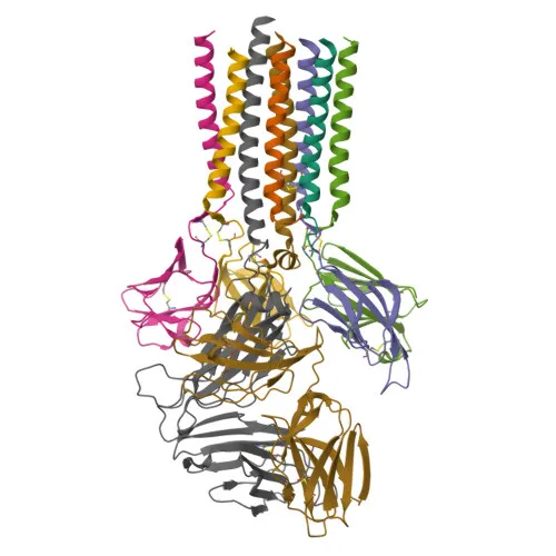
Polyclonal Antibodies
Anti-CD3D Rabbit Polyclonal Antibody for WB, IHC-P, IF/ICC, ELISA - P04234
Item Number : CM0018477
Price varies based on specs and customizations
- Application
- WB, IHC-P, IF/ICC, ELISA
- Cross Reactivity
- Human, Rat
- Protein Weight
- 19kDa

Shipping:
Contact us to get shipping details Enjoy On-time Dispatch Guarantee.
Why Choose Us
Easy ordering process, quality products, and dedicated support for your business success.
Core Product Specifications and Parameters
| Parameter | Specification |
|---|---|
| Product Name | CD3D Rabbit pAb |
| Remarks/Alias | T3D, IMD19, CD3DELTA, CD3-DELTA, CD3D |
| Species | Human |
| GeneID (Human) | 915 |
| GeneID | 915 |
| Immunogen | Recombinant fusion protein containing a sequence corresponding to amino acids 22-105 of human CD3D (NP_000723.1) |
| Source | Rabbit |
| Category | Polyclonal Antibodies |
| Application | WB, IHC-P, IF/ICC, ELISA |
| Cross Reactivity | Human, Rat |
| SWISS | P04234 |
| Protein Weight | 19kDa |
| Shipping | Ice bag |
Biological Background: CD3D Function and Localization
- CD3D encodes the T-cell surface glycoprotein CD3 delta chain, also known as the T-cell receptor T3 delta chain. It is a single-pass type I membrane protein and a key component of the TCR-CD3 complex.
- As part of the TCR-CD3 complex, CD3D is essential for adaptive immunity. Upon antigen recognition by the TCR, the CD3 chains transmit activation signals across the cell membrane via their immunoreceptor tyrosine-based activation motifs (ITAMs). These ITAMs are phosphorylated by Src family kinases LCK and FYN, initiating downstream signaling cascades (PubMed:2470098). Related references: PMID:2470098
- CD3D plays a critical role in thymocyte differentiation by ensuring correct TCR-CD3 complex assembly and surface expression. It also interacts with the CD4 and CD8 co-receptors, establishing a functional link between the TCR and these co-receptors, which is necessary for positive selection of CD4+ and CD8+ T cells (PubMed:12215456). Related references: PMID:12215456
- Subcellularly, CD3D is localized to the cell membrane, consistent with its function as a transmembrane signaling receptor.
- CD3D is predominantly expressed on T lymphocytes together with other TCR-CD3 components. It is also present on fetal NK cells, indicating a broader role in lymphocyte development.
- Post-translational modifications include phosphorylation of ITAMs, N-linked glycosylation, and disulfide bond formation, all critical for its structure and signaling functions.
Experimental Guidance and Technical Tips
- For Western blotting, consider that the immunogen corresponds to amino acids 22–105 of the extracellular region; a band near 19 kDa is expected. Use a suitable positive control such as Jurkat cell lysate.
- In IHC-P, optimize antigen retrieval conditions and validate staining with known T-cell–rich tissues (e.g., tonsil, lymph node).
- For IF/ICC, permeabilization is required to detect CD3D, and co-staining with CD4/CD8 can help confirm T-cell specificity. Validate the protocol in your sample system.
- In ELISA, use the antibody as capture or detection reagent after confirming its performance with recombinant CD3D or cell lysates; cross-reactivity with rat CD3D allows multi-species assay design.
- As a rabbit polyclonal antibody, it may recognize multiple epitopes; therefore, confirm specificity in each application using appropriate knockout or knockdown controls.
CamelBio: Your One-Stop Sourcing Bridge
CamelBio is a dedicated IVD raw-material supplier, offering diagnostic manufacturers and research institutes comprehensive access to validated antibodies, matched antibody pairs, and bulk ancillary reagents. This anti-CD3D polyclonal antibody supports immunology and T-cell research applications, and we can also assist in sourcing rare-target raw materials and developing custom immunoassay components. Contact us to streamline your supply chain from concept to clinic.
Product Datasheet
Anti-CD3D Rabbit Polyclonal Antibody for WB, IHC-P, IF/ICC, ELISA - P04234
REQUEST A QUOTE
Our professional team will reply to you within one business day. Please feel free to contact us!