Polyclonal Antibodies
Anti-CD324/E-Cadherin Polyclonal Antibody for WB, ELISA - P09803
Item Number : CM0023352
Price varies based on specs and customizations
- Application
- WB, ELISA
- Cross Reactivity
- Human, Mouse
- Protein Weight
- 98kDa

Shipping:
Contact us to get shipping details Enjoy On-time Dispatch Guarantee.
Why Choose Us
Easy ordering process, quality products, and dedicated support for your business success.
Core Product Specifications and Parameters
| Parameter | Value |
|---|---|
| Product Name | CD324/E-Cadherin Rabbit pAb |
| Remarks/Alias | CDH1; Arc-1; CD324; CDHE; ECAD; LCAM; UVO; AA960649; ARC-1; E-cad; Ecad; L-CAM; Um; cadherin-1; CD324/E-Cadherin |
| Species | Mouse |
| GeneID (Human) | 999 |
| GeneID | 12550 |
| Immunogen | Recombinant fusion protein containing a sequence corresponding to amino acids 157-369 of mouse CD324/E-Cadherin (NP_033994.1). |
| Source | Rabbit |
| Category | Polyclonal Antibodies |
| Application | WB, ELISA |
| Cross Reactivity | Human, Mouse |
| SWISS | P09803 |
| Protein Weight | 98kDa |
| Shipping | Ice bag |
Biological Background: Cadherin-1 Function and Localization
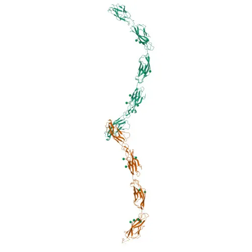
- Cadherin-1 (CDH1, E-cadherin) is a calcium-dependent cell adhesion protein that mediates homophilic interactions, contributing to cell sorting and the maintenance of epithelial tissue integrity (PubMed:11976333). Related references: PMID:11976333
- It regulates epithelial cell mobility and proliferation and functions as a potent tumor invasion suppressor.
- CDH1 promotes the organization of radial actin fibers at cell junctions via interaction with angiomotin-like 2 (AMOTL2) and facilitates early desmosome assembly by recruiting desmoglein-2 (DSG2) and desmoplakin (DSP).
- The protein localizes primarily to adherens junctions and desmosomes at the cell membrane, but is also found in endosomes, the trans-Golgi network, and the cytoplasm.
- CDH1 is widely expressed in non-neural epithelial tissues; at the protein level, it is detected in epidermal keratinocytes from birth, inner and outer pillar cells of the organ of Corti, and granuloma macrophages (PubMed:30639848, PubMed:27760340, PubMed:18079750, PubMed:27375112, PubMed:30660666). Related references: PMID:30639848 PMID:27760340 PMID:18079750 PMID:27375112 PMID:30660666
- A C-terminal proteolytic fragment, E-Cad/CTF2, has been shown to promote non-amyloidogenic degradation of amyloid-beta precursors and to inhibit the production of APP C99 and C83 peptides.
- Mouse CdH1 does not serve as a receptor for Listeria monocytogenes internalin A (InlA); a single surface residue change can confer susceptibility to bacterial uptake.
- Post-translational modifications include cleavage at basic residues, disulfide bond formation, glycosylation, phosphorylation, and ubiquitination.
Experimental Guidance and Technical Tips
- The immunogen spans amino acids 157–369 within the extracellular cadherin repeat domains; the polyclonal nature may yield recognition of multiple epitopes and detection of both full-length and processed E-cadherin forms.
- In Western blotting, expect a predominant band near 98 kDa; consider using epithelial cell lines such as MCF-7 (human) or 4T1 (mouse) as positive controls, and validate with appropriate loading controls.
- For ELISA, titrate the antibody to determine the optimal working dilution, and include calcium ions in assay buffers to preserve native E-cadherin conformation.
- Given the reported cross-reactivity with human and mouse, verify specificity in your particular sample type; endogenous expression levels can vary among different epithelial tissues.
- Avoid repeated freeze-thaw cycles; aliquot and store at recommended temperatures upon receipt that the shipping condition (ice bag) implies short-term stability during transit.
CamelBio: Your One-Stop Sourcing Bridge
CamelBio serves as a comprehensive raw-material partner for diagnostic manufacturers and research institutions, covering requirements from discovery to clinical application. This anti-CD324/E-Cadherin polyclonal antibody is one of many cell adhesion and epithelial biology reagents we supply, including validated antibody pairs, optimized monoclonal antibodies, and ancillary bulk reagents. Our sourcing expertise also extends to rare-target antibodies and custom development, ensuring you have a reliable single source for your IVD material needs.
REQUEST A QUOTE
Our professional team will reply to you within one business day. Please feel free to contact us!