Polyclonal Antibodies
Anti-CD27 Polyclonal Antibody for WB, ELISA - P26842
Item Number : CM0018396
Price varies based on specs and customizations
- Application
- WB, ELISA
- Cross Reactivity
- Mouse
- Protein Weight
- 29kDa

Shipping:
Contact us to get shipping details Enjoy On-time Dispatch Guarantee.
Why Choose Us
Easy ordering process, quality products, and dedicated support for your business success.
Core Product Specifications and Parameters
| Parameter | Value |
|---|---|
| Product Name | CD27 Rabbit pAb |
| Remarks/Alias | T14; S152; Tp55; TNFRSF7; S152. LPFS2; CD27 |
| Species | Human |
| GeneID (Human) | 939 |
| GeneID | 939 |
| Immunogen | Recombinant Protein |
| Source | Rabbit |
| Category | Polyclonal Antibodies |
| Application | WB, ELISA |
| Cross Reactivity | Mouse |
| SWISS | P26842 |
| Protein Weight | 29kDa |
| Shipping | Ice bag |
Biological Background: CD27 Function and Localization
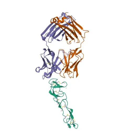
- CD27 antigen (aliases: T14, TNFRSF7, Tp55) is a member of the tumor necrosis factor receptor superfamily, functioning as a costimulatory immune-checkpoint receptor on the surface of T-cells, NK-cells, and B-cells.
- Its primary ligand is CD70 (CD27L), and the CD70-CD27 pathway drives antigen-specific T-cell activation and expansion, providing immune surveillance of B-cells. Related references: PMID:28011863
- Mechanistically, CD70 binding activates the TRAF2-PTPN6 axis, inhibiting LCK phosphorylation to promote T-cell memory adaptations. Related references: PMID:38354704
- Additionally, CD27 engagement on early progenitor cells delivers a negative feedback signal to leukocyte differentiation, modulating hematopoiesis, and it negatively regulates Th2 lymphocyte function in adipose tissue.
- The protein is a single-pass transmembrane receptor localized exclusively at the cell membrane and contains a signal peptide, extracellular TNFR-like cysteine-rich domains, a transmembrane helix, and a cytoplasmic tail.
- Key post‑translational modifications include N‑linked glycosylation and phosphorylation; the receptor is also characterized by disulfide bonds that stabilize its extracellular structure.
- CD27 is broadly expressed on most T‑lymphocytes, and its signaling is implicated in apoptosis, disease variants of lymphoproliferative syndromes, and hematopoietic regulation.
Experimental Guidance and Technical Tips
- The immunogen corresponds to the extracellular domain (amino acids 20–191) of human CD27, suggesting the antibody likely recognizes epitopes within the ligand-binding region; consider performing epitope mapping if precise binding site information is needed.
- For Western blot, the predicted molecular weight is 29 kDa; however, due to glycosylation, the observed band may migrate at a higher apparent molecular weight. Validate under reducing and non‑reducing conditions as appropriate.
- The antibody is reported to cross‑react with mouse CD27, making it a potential tool for comparative studies; always verify cross‑reactivity in the relevant experimental system.
- ELISA applications may benefit from pairing this polyclonal antibody with a monoclonal detection antibody in a sandwich format; optimize coating and detection conditions empirically.
- As a polyclonal preparation, lot‑to‑lot consistency should be monitored in quantitative assays; consider using a recombinant monoclonal antibody if stringent reproducibility is required.
CamelBio: Your One-Stop Sourcing Bridge
CamelBio offers diagnostic manufacturers, contract research organizations, and academic laboratories a streamlined channel to procure validated IVD raw materials like this anti‑CD27 polyclonal antibody. Our portfolio spans a wide range of immune‑checkpoint and T‑cell targets, including optimized monoclonal/polyclonal antibodies, matched antibody pairs, and bulk ancillary reagents. Through expert consultation and global supply networks, we help accelerate your CD27‑based assay development and rare‑target sourcing needs.
REQUEST A QUOTE
Our professional team will reply to you within one business day. Please feel free to contact us!