Polyclonal Antibodies
Anti-CD200R1 Rabbit Polyclonal Antibody for WB, ELISA - Q8TD46
Item Number : CM0021025
Price varies based on specs and customizations
- Application
- WB, ELISA
- Cross Reactivity
- Human
- Protein Weight
- 37kDa

Shipping:
Contact us to get shipping details Enjoy On-time Dispatch Guarantee.
Why Choose Us
Easy ordering process, quality products, and dedicated support for your business success.
Core Product Specifications and Parameters
| Parameter | Value |
|---|---|
| Product Name | CD200R1 Rabbit pAb |
| Remarks/Alias | OX2R; MOX2R; CD200R; HCRTR2; CD200R1 |
| Species | Human |
| Gene ID (Human) | 131450 |
| Gene ID | 131450 |
| Immunogen | Recombinant fusion protein containing a sequence corresponding to amino acids 29-240 of human CD200R1 (NP_740750.1) |
| Source | Rabbit |
| Category | Polyclonal Antibodies |
| Application | WB, ELISA |
| Cross Reactivity | Human |
| SWISS | Q8TD46 |
| Protein Weight | 37kDa |
| Shipping | Ice bag |
Biological Background: CD200R1 Function and Localization
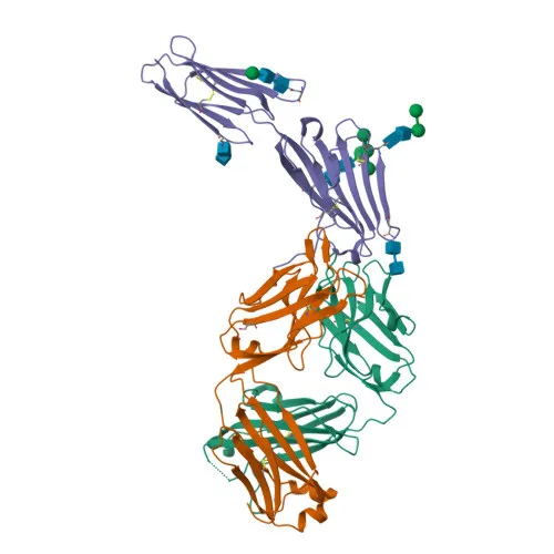
- Cell surface glycoprotein CD200 receptor 1 (CD200R1), also known as OX2 receptor 1, MOX2R, and HCRTR2, is a member of the immunoglobulin superfamily and functions as an inhibitory receptor.
- Binds the CD200/OX2 ligand to limit inflammation by suppressing the expression of pro-inflammatory molecules such as TNF, interferons, and inducible nitric oxide synthase (iNOS).
- Interacts with the HHV-8 K14 viral CD200 homolog with similar affinity and kinetics as the endogenous ligand, implicating a role in host-virus interaction.
- Alternative splicing produces both membrane-bound and secreted isoforms; the receptor is primarily localized to the cell membrane but can be released as a soluble form.
- Tissue expression is predominantly in hematopoietic cells: granulocytes, monocytes, most T-cells, neutrophils, basophils, and subsets of NK, NKT, and B-cells; also found in bone marrow, lymph nodes, spleen, lung, liver, spinal cord, kidney, monocyte-derived dendritic cells, and mast cells.
- Post-translational modifications include glycosylation and disulfide bond formation, consistent with a cell surface glycoprotein.
- The protein contains a signal peptide, a single transmembrane helix, and extracellular immunoglobulin-like domains.
Experimental Guidance and Technical Tips
- The immunogen corresponds to the extracellular domain (aa 29-240), which is accessible on the cell surface and in secreted forms; this region is suitable for detecting both membrane-bound and soluble CD200R1.
- For Western blotting, the predicted molecular weight is approximately 37 kDa, but glycosylation may cause a shift; consider using endoglycosidase treatment to confirm specificity.
- In ELISA applications, coating with recombinant CD200R1 protein or capture from biological fluids may require optimization of buffer conditions and detection antibodies.
- Validate specificity in relevant human immune cell lines (e.g., Jurkat, THP-1) or primary leukocyte lysates, as expression is restricted to hematopoietic lineages.
CamelBio: Your One-Stop Sourcing Bridge
CamelBio is a dedicated B2B supplier of IVD raw materials, offering validated antibody pairs, monoclonal and polyclonal antibodies, bulk ancillary reagents, and specialized rare-target sourcing for diagnostic manufacturers and research laboratories. CD200R1, as an immune checkpoint receptor, is a promising biomarker in inflammation and viral pathogenesis studies, and CamelBio’s comprehensive sourcing capabilities help meet your assay development needs from early-stage research to clinical application.
REQUEST A QUOTE
Our professional team will reply to you within one business day. Please feel free to contact us!