Polyclonal Antibodies
Anti-CD1D Rabbit Polyclonal Antibody for WB, IF/ICC, ELISA - P15813
Item Number : CM0019312
Price varies based on specs and customizations
- Application
- WB, IF/ICC, ELISA
- Cross Reactivity
- Human, Mouse
- Protein Weight
- 38kDa

Shipping:
Contact us to get shipping details Enjoy On-time Dispatch Guarantee.
Why Choose Us
Easy ordering process, quality products, and dedicated support for your business success.
Core Product Specifications and Parameters
| Parameter | Value |
|---|---|
| Product Name | CD1D Rabbit pAb |
| Remarks/Alias | R3; CD1A; R3G1; CD1D |
| Species | Human |
| GeneID (Human) | 912 |
| GeneID | 912 |
| Immunogen | Recombinant fusion protein containing a sequence corresponding to amino acids 20-190 of human CD1D (NP_001757.1). |
| Source | Rabbit |
| Category | Polyclonal Antibodies |
| Application | WB, IF/ICC, ELISA |
| Cross Reactivity | Human, Mouse |
| SWISS | P15813 |
| Protein Weight | 38kDa |
| Shipping | Ice bag |
Biological Background: CD1D Function and Localization
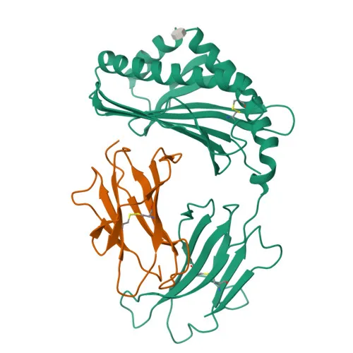
- CD1D (R3G1) is an antigen-presenting glycoprotein of the CD1 family, primarily involved in presenting lipid antigens to natural killer T (NKT) cells.
- It binds self and non-self glycolipids within its hydrophobic binding groove and presents them to T-cell receptors on NKT cells, bridging innate and adaptive immunity.
- CD1D is expressed on cortical thymocytes, certain T-cell leukemias, and in a variety of other tissues, supporting its role in immune surveillance.
- The protein localizes to the cell membrane, basolateral cell membrane, endosome membrane, lysosome membrane, and endoplasmic reticulum membrane, reflecting its trafficking through the endocytic pathway for antigen loading.
- Post‑translational modifications include glycosylation and disulfide bond formation, contributing to its structural stability and ligand‑binding function.
- The predicted molecular weight of CD1D is approximately 37.7 kDa, though glycosylation may result in a higher apparent molecular weight (observed at 38 kDa in some systems).
Experimental Guidance and Technical Tips
- For Western blot, consider loading appropriate positive controls such as lymphoid cell lines (e.g., Jurkat) and using reducing loading buffer to resolve the ~38 kDa band.
- When performing immunofluorescence, permeabilize cells with 0.1% Triton X-100 to detect intracellular pools of CD1D.
- Since the antibody cross-reacts with mouse, validate its specificity in your model system by using CD1D knockout or knockdown controls.
- ELISA applications may require optimization of coating antigen and detection conditions.
- This polyclonal antibody was raised against the extracellular domain (aa 20-190); thus, it may detect both membrane-bound and soluble forms of CD1D.
CamelBio: Your One-Stop Sourcing Bridge
CamelBio is your one-stop sourcing partner for IVD raw materials, supplying validated antibody pairs, optimized monoclonal/polyclonal antibodies, bulk ancillary reagents, and rare target raw-material sourcing. This CD1D antibody supports research into innate immunity and glycolipid antigen presentation. Reach out for technical services and tailored procurement solutions from concept to clinic.
REQUEST A QUOTE
Our professional team will reply to you within one business day. Please feel free to contact us!