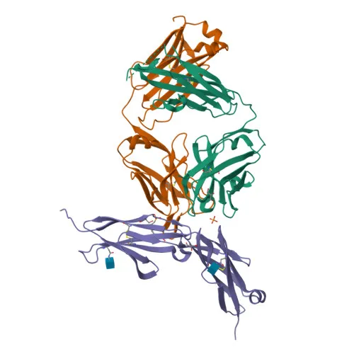
Monoclonal Antibodies
Anti-CD146/MCAM Rabbit Monoclonal Antibody for WB, IHC-P, IF/ICC - P43121
Item Number : CM0010982
Price varies based on specs and customizations
- Application
- WB, IF/ICC, IHC-P, ELISA
- Cross Reactivity
- Human
- Protein Weight
- 72 kDa

Shipping:
Contact us to get shipping details Enjoy On-time Dispatch Guarantee.
Why Choose Us
Easy ordering process, quality products, and dedicated support for your business success.
Core Product Specifications and Parameters
| Parameter | Value |
|---|---|
| Product Name | CD146/MCAM Rabbit mAb |
| Remarks/Alias | CD146; MUC18; HEMCAM; METCAM; MelCAM |
| Species | Human |
| GeneID (human) | 4162 |
| GeneID | 4162 |
| Immunogen | Recombinant fusion protein containing a sequence corresponding to amino acids 24-559 of human CD146/MCAM (NP_006491.2) |
| Source | Rabbit |
| Category | Monoclonal Antibodies |
| Application | WB, IF/ICC, IHC-P, ELISA |
| Cross Reactivity | Human |
| SWISS | P43121 |
| Protein Weight | 72 kDa |
| Shipping | Ice bag |
Biological Background: CD146/MCAM Function and Localization
- CD146/MCAM (also known as MUC18, MelCAM, HEMCAM) is a cell surface glycoprotein encoded by the MCAM gene, belonging to the immunoglobulin superfamily of adhesion molecules.
- It mediates calcium-independent cell-cell adhesion and is critical for maintaining endothelial monolayer integrity at intercellular junctions.
- Its expression on melanoma cells facilitates interactions with vascular endothelium, promoting hematogenous dissemination and metastasis.
- During embryonic development, CD146/MCAM may function as an adhesion molecule in migrating neural crest cells.
- Ligand engagement triggers tyrosine phosphorylation of FYN and PTK2/FAK1, along with a transient rise in intracellular calcium, indicating a role in signal transduction.
- In normal adult tissues, CD146/MCAM expression is largely restricted to vascular smooth muscle and endothelial cells.
- The protein is highly expressed in advanced primary and metastatic melanomas but rarely detected in benign nevi, correlating with tumor progression.
- Post-translational modifications include phosphorylation and N-linked glycosylation, and the protein contains disulfide-linked immunoglobulin-like repeats and a transmembrane domain.
Experimental Guidance and Technical Tips
- The immunogen corresponds to the extracellular domain (aa 24-559) of human CD146/MCAM, predicting detection of the full-length protein and any soluble shed forms in appropriate samples.
- For Western blot, the predicted band is ~72 kDa; however, due to glycosylation, the observed molecular weight may be higher. Consider using denaturing and reducing conditions.
- In immunofluorescence (IF/ICC), membrane localization should be prominent; include appropriate permeabilization if intracellular epitopes are to be accessed, though the extracellular domain targeting may allow live-cell staining.
- For immunohistochemistry (IHC-P), antigen retrieval methods and optimization may be required; expression is expected in vascular endothelium and melanoma tissue.
- While ELISA is listed as an application, pair with a suitable capture/detection antibody; end-users should validate the antibody in their specific assay format.
- Cross-reactivity has been validated for human; for other species, perform sequence homology analysis and experimental verification.
CamelBio: Your One-Stop Sourcing Bridge
CamelBio serves as a strategic one-stop sourcing partner for diagnostic manufacturers and research laboratories requiring high-quality IVD raw materials. Our portfolio includes this CD146/MCAM rabbit monoclonal antibody, tailored for oncology and vascular biology applications, alongside complementary products such as antibody pairs, conjugated detection reagents, and bulk ancillary reagents. With supply chain expertise in rare-target raw materials, CamelBio supports your workflow from concept to clinical implementation.
REQUEST A QUOTE
Our professional team will reply to you within one business day. Please feel free to contact us!