Monoclonal Antibodies
Anti-CD109 Monoclonal Antibody for WB, ELISA - Q6YHK3
Item Number : CM0007329
Price varies based on specs and customizations
- Application
- WB, ELISA
- Cross Reactivity
- Human
- Protein Weight
- 74kDa/152kDa/159kDa/161kDa

Shipping:
Contact us to get shipping details Enjoy On-time Dispatch Guarantee.
Why Choose Us
Easy ordering process, quality products, and dedicated support for your business success.
Core Product Specifications and Parameters
| Parameter | Value |
|---|---|
| Product Name | CD109 Rabbit mAb |
| Remarks/Alias | CD109; CPAMD7; p180; r150; CD109 antigen |
| Species | Human |
| GeneID (Human) | 1024 |
| GeneID | 135228 |
| Immunogen | Recombinant protein | Recombinant fusion protein containing a sequence corresponding to amino acids 223-462 of human CD109. (NP_598000.2) |
| Source | Rabbit |
| Category | Monoclonal Antibodies |
| Application | WB, ELISA |
| Cross Reactivity | Human |
| SWISS | Q6YHK3 |
| Protein Weight | 74kDa/152kDa/159kDa/161kDa |
| Shipping | Ice bag |
Biological Background: CD109 Function and Localization
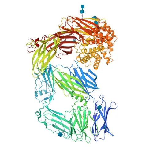
- CD109 is a GPI-anchored cell membrane glycoprotein belonging to the alpha-2-macroglobulin family, also known as 150 kDa TGF-beta-1-binding protein, C3 and PZP-like alpha-2-macroglobulin domain-containing protein 7, platelet-specific Gov antigen, p180, and r150.
- Encoded by the CD109 (CPAMD7) gene, it is a member of the alpha-2-macroglobulin superfamily, characterized by a conserved thioester bond and protease inhibitor domains.
- CD109 is localized at the cell membrane via a glycosylphosphatidylinositol (GPI) linkage, consistent with its role in cell surface signaling and protein interactions.
- The primary function of CD109 is the negative modulation of transforming growth factor beta 1 (TGFB1) signaling, especially in keratinocytes.
- Multiple isoforms arise from alternative splicing; isoform 1 is expressed in keratinocytes and placenta, while isoform 4 is placental, with molecular weights varying from 74 kDa to 161 kDa due to isoform length and post-translational modifications.
- Post-translational modifications include glycosylation, GPI-anchor attachment, and a thioester bond, which contribute to its structural stability and functional activity.
- CD109 is widely expressed at high levels in the uterus, aorta, heart, lung, trachea, and placenta, and in fetal heart, kidney, liver, spleen, and lung. It is also found in CD34+ acute myeloid leukemia cell lines, T-cell lines, activated T-lymphoblasts, endothelial cells, and activated platelets.
Experimental Guidance and Technical Tips
- The immunogen (amino acids 223–462) targets an extracellular region, which may preserve native epitopes. Consider verifying epitope accessibility for different applications.
- For Western blotting, multiple bands may be observed due to glycosylation and isoform heterogeneity. Use both reducing and non-reducing conditions, and validate in relevant positive controls such as lysates from human keratinocytes, placenta, or platelet-rich plasma.
- In ELISA assays, this antibody can be explored as a capture or detection reagent. Pair with a suitable matched antibody or direct conjugate, and optimize using recombinant CD109 protein or platelet-derived samples.
CamelBio: Your One-Stop Sourcing Bridge
CamelBio provides diagnostic manufacturers, labs, and research institutes with one-stop access to IVD raw materials, technical services, and consulting—covering every stage from concept to clinic. Our portfolio includes validated monoclonal antibodies like this anti-CD109 clone, supporting TGF-beta signaling and platelet research. From antibody pairs and bulk ancillary reagents to rare target sourcing, we streamline procurement to accelerate your diagnostic development.
REQUEST A QUOTE
Our professional team will reply to you within one business day. Please feel free to contact us!