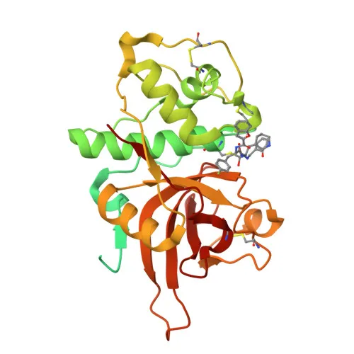
Monoclonal Antibodies
Anti-Cathepsin L/V/K/H Rabbit Monoclonal Antibody for WB, IHC-P, ELISA - P07711/O60911/P43235/P09668
Item Number : CM0008338
Price varies based on specs and customizations
- Application
- WB, IHC-P, ELISA
- Cross Reactivity
- Human, Mouse, Rat
- Protein Weight
- 38kDa

Shipping:
Contact us to get shipping details Enjoy On-time Dispatch Guarantee.
Why Choose Us
Easy ordering process, quality products, and dedicated support for your business success.
Core Product Specifications and Parameters
| Parameter | Value |
|---|---|
| Product Name | Cathepsin L/V/K/H Rabbit mAb |
| Remarks/Alias | MEP; CATL; CTSL1; Cathepsin L/V/K/H |
| Species | Human |
| Gene ID (Human) | 1514 |
| Gene ID | 1514/1515/1513/1512 |
| Immunogen | Synthetic Peptide - A synthetic peptide corresponding to a sequence within amino acids 50-150 of human Cathepsin L/V/K/H (P07711). |
| Source | Rabbit |
| Category | Monoclonal Antibodies |
| Application | WB, IHC-P, ELISA |
| Cross Reactivity | Human, Mouse, Rat |
| SWISS | P07711/O60911/P43235/P09668 |
| Protein Weight | 38kDa |
| Shipping | Ice bag |
Biological Background: Cathepsin L Function and Localization
- Procathepsin L (Cathepsin L1), also known as major excreted protein (MEP), is a lysosomal cysteine protease encoded by the CTSL gene. It belongs to the peptidase C1 family and shares high homology with cathepsins V, K, and H.
- As a thiol protease, cathepsin L is essential for general protein turnover within lysosomes. It participates in diverse processes including antigen processing in thymic epithelial cells, bone remodeling, and the proteolytic release of thyroid hormone thyroxine from thyroglobulin.
- Cathepsin L degrades elastin at neutral pH and accumulates in the extracellular space of antigen-presenting cells, contributing to extracellular matrix remodeling during inflammation. It also generates the angiogenesis inhibitor endostatin from collagen XVIII. Related references: PMID:10716919
- In cells lacking TMPRSS2, cathepsin L facilitates the entry of SARS-CoV, SARS-CoV-2, and other coronaviruses by cleaving spike glycoproteins within lysosomes, enabling membrane fusion.
Related references:
PMID:16339146
PMID:18562523
PMID:32142651
PMID:16081529
PMID:26953343 - Alternative translation initiation produces isoforms lacking the signal peptide, which localize to the nucleus and cleave the CUX1 transcription factor, thereby modulating cell cycle progression. Related references: PMID:15099520
- Cathepsin L is primarily localized to lysosomes but can also be found at the apical cell membrane, in secretory vesicles (e.g., chromaffin granules), in the extracellular space, and in the nucleus under certain conditions.
- Key post-translational modifications include glycosylation and disulfide bond formation. The enzyme is synthesized as an inactive zymogen (procathepsin L) and activated by proteolytic cleavage. Its mature form migrates at approximately 38 kDa on SDS-PAGE, while the full-length precursor is around 37.6 kDa.
Experimental Guidance and Technical Tips
- The immunogen is a synthetic peptide from the conserved region (aa 50–150) of human cathepsin L, which likely accounts for cross-reactivity with cathepsins V, K, and H. When interpreting results, consider possible detection of multiple family members.
- For western blotting, a band near 38 kDa is expected under reducing conditions. Note that procathepsin L (~37.5 kDa) and its processed forms may also be visible depending on sample preparation and cellular context.
- In immunohistochemistry (IHC-P), antigen retrieval methods may be required to unmask epitopes in formalin-fixed tissues. Optimization of retrieval conditions (e.g., citrate vs. EDTA buffer, pH, and heating) is recommended.
- For ELISA, this antibody may be suitable as a capture or detection reagent. Validate in the appropriate sample matrix (e.g., cell lysates, tissue extracts, or biological fluids) and confirm specificity with recombinant protein controls.
- Given its expression in a wide range of tissues and cell types, including immune cells and epithelia, include appropriate positive and negative controls. For studies involving viral entry, note that cathepsin L acts in acidic compartments; inhibitors like leupeptin or E-64 can be used to confirm specificity.
CamelBio: Your One-Stop Sourcing Bridge
CamelBio supports diagnostic developers and research laboratories with a comprehensive portfolio of IVD raw materials, including validated antibody pairs, optimized monoclonal and polyclonal antibodies, and bulk ancillary reagents. This anti-cathepsin L/V/K/H antibody exemplifies the type of high-quality protease-targeting reagents we can source for applications in lysosomal biology, infectious disease research, and biomarker assay development. Through our one-stop platform, we help you secure rare-target raw materials and accelerate your transition from concept to clinical application.
Product Datasheet
Anti-Cathepsin L/V/K/H Rabbit Monoclonal Antibody for WB, IHC-P, ELISA - P07711/O60911/P43235/P09668
REQUEST A QUOTE
Our professional team will reply to you within one business day. Please feel free to contact us!