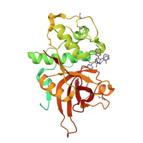
Polyclonal Antibodies
Anti-Cathepsin L/V/K/H Polyclonal Antibody for WB, ELISA - P07711
Item Number : CM0018559
Price varies based on specs and customizations
- Application
- WB, ELISA
- Cross Reactivity
- Human, Rat
- Protein Weight
- 38kDa

Shipping:
Contact us to get shipping details Enjoy On-time Dispatch Guarantee.
Why Choose Us
Easy ordering process, quality products, and dedicated support for your business success.
Core Product Specifications and Parameters
| Parameter | Value |
|---|---|
| Product Name | Cathepsin L/V/K/H Rabbit pAb |
| Remarks/Alias | MEP; CATL; CTSL1; Cathepsin L/V/K/H |
| Species | Human |
| Gene ID (Human) | 1514 |
| Gene ID | 1514 |
| Immunogen | Recombinant protein |
| Source | Rabbit |
| Category | Polyclonal Antibodies |
| Application | WB, ELISA |
| Cross Reactivity | Human, Rat |
| SWISS | P07711 |
| Protein Weight | 38kDa |
| Shipping | Ice bag |
Biological Background: Cathepsin L/V/K/H Function and Localization
- Cathepsin L, encoded by CTSL (also known as Cathepsin L1 or MEP), is a lysosomal cysteine protease synthesized as an inactive zymogen (procathepsin L) that is activated by proteolytic removal of the propeptide.
- It functions as a thiol protease critical for overall protein degradation within lysosomes, and participates in antigen processing, bone remodeling, and general protein turnover.
- In the thyroid, it solubilizes cross-linked thyroglobulin to release thyroxine (T4), and in neuroendocrine chromaffin cells, it processes proenkephalin to active enkephalin peptides.
- Secreted procathepsin L generates the angiogenesis inhibitor endostatin from collagen XVIII (COL18A1).
Related references: PMID:10716919 - It facilitates cellular entry of coronaviruses (SARS-CoV, SARS-CoV-2, EMC) and Zaire ebolavirus by proteolytically activating viral glycoproteins in lysosomes when TMPRSS2 is absent.
Related references: PMID:16339146 PMID:18562523 PMID:32142651 PMID:16081529 PMID:26953343 - Nuclear isoforms, produced via alternative translation initiation, proteolytically process the CUX1 transcription factor to regulate cell cycle progression.
Related references: PMID:15099520 - The protein localizes to lysosomes, apical cell membrane, secretory vesicles, chromaffin granules, extracellular space, and nucleus; it is annotated with keywords including alternative initiation, disulfide bond, glycoprotein, host-virus interaction, thiol protease, and zymogen.
Experimental Guidance and Technical Tips
- The immunogen covers amino acids 114–333 of human Cathepsin L, a region that shares homology with cathepsins V, K, and H; cross-reactivity with these family members should be validated in the relevant sample system.
- For Western blot, the antibody is expected to detect a band around 38 kDa (pro-form) and possibly a 25 kDa mature form; consider using appropriate positive controls (e.g., lysates from human or rat tissues/lines).
- In ELISA, the rabbit polyclonal can be paired with a mouse monoclonal against a distinct epitope for sandwich detection; optimize concentrations by checkerboard titration.
- Since the antibody reacts with human and rat, it may be suitable for cross-species studies, but performance in other species must be experimentally confirmed.
- Dilutions and incubation conditions should be optimized empirically; no guaranteed working dilutions are provided.
CamelBio: Your One-Stop Sourcing Bridge
CamelBio provides diagnostic manufacturers and research labs with a reliable sourcing channel for IVD antibodies and reagents. Complement your Cathepsin L/V/K/H studies with validated antibody pairs, recombinant proteins, and bulk ancillary reagents. Our team assists in procuring rare-target raw materials, accelerating your assay development from concept to clinic.
REQUEST A QUOTE
Our professional team will reply to you within one business day. Please feel free to contact us!