Monoclonal Antibodies
Anti-Cathepsin H Rabbit Monoclonal Antibody for WB, ELISA - P09668
Item Number : CM0007093
Price varies based on specs and customizations
- Application
- WB, ELISA
- Cross Reactivity
- Human, Mouse
- Protein Weight
- 37kDa

Shipping:
Contact us to get shipping details Enjoy On-time Dispatch Guarantee.
Why Choose Us
Easy ordering process, quality products, and dedicated support for your business success.
Core Product Specifications and Parameters
| Parameter | Value |
|---|---|
| Product Name | Cathepsin H Rabbit mAb |
| Remarks/Alias | ACC4; ACC5; CPSB; ACC-4; ACC-5; Cathepsin H |
| Species | Human |
| Gene ID | 1512 |
| Immunogen | Recombinant fusion protein containing a sequence corresponding to amino acids 150-250 of human Cathepsin H (NP_004381.2). |
| Source | Rabbit |
| Category | Monoclonal Antibodies |
| Application | WB, ELISA |
| Cross Reactivity | Human, Mouse |
| SWISS | P09668 |
| Protein Weight | 37kDa |
| Shipping | Ice bag |
Biological Background: Cathepsin H Function and Localization
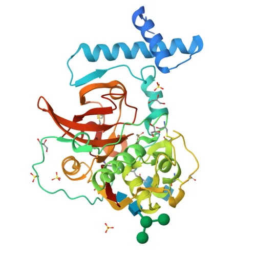
- Cathepsin H is encoded by the CTSH gene (also known as CPSB) and exists as a pro-enzyme (pro-cathepsin H) that is proteolytically processed into its mature, active form.
- It functions as a lysosomal cysteine protease, playing a key role in the overall degradation of proteins within the lysosome.
- Cathepsin H belongs to the papain family of cysteine proteases and exhibits both endopeptidase and aminopeptidase activities.
- The protein is characterized as a thiol protease, relying on a cysteine residue in its active site for catalytic activity.
- It is synthesized as a zymogen (pro-cathepsin H) and undergoes post-translational modifications including glycosylation and disulfide bond formation, which are critical for proper folding and stability.
- Cathepsin H is predominantly localized to the lysosome, consistent with its role in intracellular protein turnover.
- As evidenced by keywords such as "Glycoprotein" and "Disulfide bond," its structure includes carbohydrate modifications and stabilizing disulfide linkages.
Experimental Guidance and Technical Tips
- For Western blot, target band detection around 37 kDa corresponding to pro-cathepsin H; note that processed forms may appear at lower molecular weights.
- Given the recombinant immunogen spanning a conserved region, validate antibody specificity in relevant human and mouse tissue or cell lysates to confirm expected reactivity.
- For ELISA applications, consider pairing this monoclonal antibody with a suitable detection reagent; assess optimal capture and detection conditions empirically.
- Since cathepsin H contains disulfide bonds, use reducing sample buffer for SDS-PAGE to ensure complete denaturation.
- The antibody's reactivity with mouse samples allows cross-species validation in preclinical models; nonetheless, confirm consistent performance prior to study initiation.
CamelBio: Your One-Stop Sourcing Bridge
CamelBio is your trusted one-stop partner for IVD raw material procurement, offering validated antibody pairs, optimized monoclonal/polyclonal antibodies, and bulk ancillary reagents. This anti-Cathepsin H antibody supports research in lysosomal degradation and protease-related disorders, and we can assist with sourcing additional rare-target raw materials to streamline your assay development. From concept to clinic, CamelBio bridges the gap with comprehensive supply solutions tailored to diagnostic innovators.
REQUEST A QUOTE
Our professional team will reply to you within one business day. Please feel free to contact us!