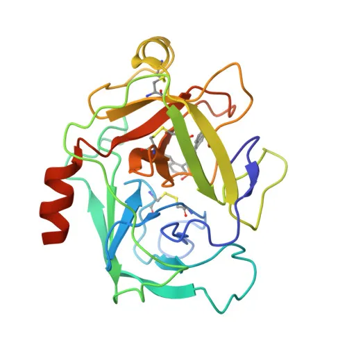
Polyclonal Antibodies
Anti-Cathepsin G Rabbit Polyclonal Antibody for WB, IHC-P, ELISA - P08311
Item Number : CM0014155
Price varies based on specs and customizations
- Application
- WB, IHC-P, ELISA
- Cross Reactivity
- Human, Mouse, Rat
- Protein Weight
- 29kDa

Shipping:
Contact us to get shipping details Enjoy On-time Dispatch Guarantee.
Why Choose Us
Easy ordering process, quality products, and dedicated support for your business success.
Core Product Specifications and Parameters
| Parameter | Value |
|---|---|
| Product Name | Cathepsin G Rabbit pAb |
| Remarks/Alias | CG; CATG; Cathepsin G |
| Species | Human |
| GeneID (Human) | 1511 |
| GeneID | 1511 |
| Immunogen | Recombinant fusion protein containing a sequence corresponding to amino acids 75-190 of human Cathepsin G (NP_001902.1). |
| Source | Rabbit |
| Category | Polyclonal Antibodies |
| Application | WB, IHC-P, ELISA |
| Cross Reactivity | Human, Mouse, Rat |
| SWISS | P08311 |
| Protein Weight | 29kDa |
| Shipping | Ice bag |
Biological Background: Cathepsin G Function and Localization
-
Cathepsin G (CTSG) is a serine protease with trypsin- and chymotrypsin-like specificity, preferentially cleaving after Phe, Tyr, Trp, and Leu at the P1 position and showing a preference for negatively charged residues at P2′ (PubMed:29652924, PubMed:8194606).
Related references: PMID:29652924 PMID:8194606 -
It exhibits potent antimicrobial activity against Gram‑negative (N. gonorrhoeae, P. aeruginosa) and Gram‑positive (L. monocytogenes, S. aureus) bacteria, as well as M. tuberculosis, largely independent of its protease function (PubMed:2116408, PubMed:2117044, PubMed:1937776, PubMed:15385470).
Related references: PMID:2116408 PMID:2117044 PMID:1937776 PMID:15385470 -
Cathepsin G modulates adaptive and innate immunity by binding to and stimulating B cells, CD4⁺ and CD8⁺ T cells, and enhancing NK cell cytotoxicity through its enzymatic activity (PubMed:7842483, PubMed:9000539, PubMed:9536127).
Related references: PMID:7842483 PMID:9000539 PMID:9536127 -
The protease is required for platelet recruitment and activation mediated by the F2RL3/PAR4 receptor; it also directly induces platelet aggregation, an effect strongly potentiated by neutrophil elastase (ELANE) (PubMed:10702240, PubMed:3390156, PubMed:25211214, PubMed:9111081).
Related references: PMID:10702240 PMID:3390156 PMID:25211214 PMID:9111081 -
It cleaves a wide range of physiological substrates including complement C3, thrombin receptor F2R/PAR1 (acting as agonist or inhibitor depending on the site), coagulation factors F8 and F10, pro‑inflammatory cytokines IL36G and IL33, and leukocyte surface protein CD43 (PubMed:1861080, PubMed:7744748, PubMed:18217133, PubMed:8920993, PubMed:30804664, PubMed:22307629, PubMed:18586676).
Related references: PMID:1861080 PMID:7744748 PMID:18217133 PMID:8920993 PMID:30804664 PMID:22307629 PMID:18586676 -
The protein is found in multiple subcellular compartments, including cytoplasmic granules of neutrophils, secreted extracellularly, and also detected in the cell membrane, cytoplasm, cytosol, lysosome, and nucleus.
-
Tissue expression is predominantly in neutrophils (at protein level) and to a lesser extent in B cells (PubMed:3799965, PubMed:15100291).
Related references: PMID:3799965 PMID:15100291 -
Cathepsin G is a glycoprotein containing intramolecular disulfide bonds; it is synthesized as an inactive zymogen and proteolytically processed to the mature enzyme.
Experimental Guidance and Technical Tips
- For Western blotting (WB), the antibody is expected to detect a band near 29 kDa corresponding to mature cathepsin G; pro‑forms or differentially glycosylated variants may also be observed. We recommend optimizing the sample preparation and loading concentrations in your specific model system.
- In immunohistochemistry (IHC‑P), antigen retrieval by heat‑mediated methods (e.g., citrate or EDTA buffer, pH 6.0 or 9.0) may improve staining. Validate the antibody on positive control tissues known to express cathepsin G (e.g., human tonsil, spleen) alongside negative controls.
- For ELISA, the polyclonal nature can provide high sensitivity, but it is crucial to assess lot‑to‑lot consistency and potential cross‑reactivity. Pairing with a suitable capture antibody or using it as a detection reagent are both viable strategies.
- The immunogen lies within the catalytic domain (aa 75–190), suggesting the antibody likely recognizes both the pro‑ and mature enzyme; thus, care should be taken when interpreting data from systems where pro‑cathepsin G is abundant.
CamelBio: Your One‑Stop Sourcing Bridge
CamelBio serves as a comprehensive, one‑stop sourcing partner for IVD raw materials, supporting diagnostic manufacturers and research laboratories from concept to clinic. For cathepsin G‑related projects, we provide validated anti‑cathepsin G rabbit polyclonal antibodies, complementary monoclonal antibodies, bulk ancillary reagents, and custom rare‑target sourcing to streamline your assay development and immunodetection workflows. Whether you need matched antibody pairs, optimized conjugates, or scalable supply, our technical team stands ready to help you accelerate your product pipeline.
Product Datasheet
Anti-Cathepsin G Rabbit Polyclonal Antibody for WB, IHC-P, ELISA - P08311
REQUEST A QUOTE
Our professional team will reply to you within one business day. Please feel free to contact us!