Monoclonal Antibodies
Anti-Cathepsin D Rabbit Monoclonal Antibody for WB, IHC-P, ELISA - P07339
Item Number : CM0002899
Price varies based on specs and customizations
- Application
- WB, IHC-P, ELISA
- Cross Reactivity
- Human, Mouse
- Protein Weight
- 45kDa

Shipping:
Contact us to get shipping details Enjoy On-time Dispatch Guarantee.
Why Choose Us
Easy ordering process, quality products, and dedicated support for your business success.
Core Product Specifications and Parameters
| Parameter | Value |
|---|---|
| Product Name | Cathepsin D Rabbit mAb |
| Remarks/Alias | CPSD; CLN10; HEL-S-130P; Cathepsin D |
| Species | Human |
| Gene ID (Human) | 1509 |
| Gene ID | 1509 |
| Immunogen | Synthetic Peptide |
| Source | Rabbit |
| Category | Monoclonal Antibodies |
| Application | WB, IHC-P, ELISA |
| Cross Reactivity | Human, Mouse |
| SWISS | P07339 |
| Protein Weight | 45kDa |
| Shipping | Ice bag |
Biological Background: Cathepsin D Function and Localization
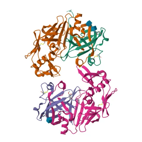
- Cathepsin D is an aspartyl protease encoded by the CTSD gene, initially produced as an inactive zymogen that undergoes proteolytic processing to its mature form.
- It functions as a major acid protease responsible for intracellular protein turnover and plays a role in APP processing after activation by ADAM30, promoting APP degradation. Related references: PMID:27333034
- The protease is implicated in the pathogenesis of several diseases, including breast cancer and possibly Alzheimer’s disease.
- At the subcellular level, Cathepsin D localizes predominantly to lysosomes, but is also found in melanosomes and can be secreted into the extracellular space.
- Tissue expression is notable in the aorta extracellular space and liver, as demonstrated at the protein level. Related references: PMID:20551380 PMID:1426530
- Post-translational modifications include N-linked glycosylation and formation of disulfide bonds essential for its structure; the protein is a member of the peptidase A1 family.
Experimental Guidance and Technical Tips
- The immunogen corresponds to a C‑terminal region (amino acids 313‑412) of human Cathepsin D, which may recognize both pro‑ and mature forms; consider this when analyzing processed variants.
- For Western blotting, a band around 45 kDa is predicted, but Cathepsin D undergoes processing that can yield multiple species; validate the observed pattern in your specific sample system.
- In IHC‑P, staining intensity and localization can vary with fixation and antigen retrieval conditions; optimization is recommended for each tissue type.
- For ELISA applications, ensure that the coating antigen contains the peptide sequence used for immunization to favor optimal antibody capture.
CamelBio: Your One-Stop Sourcing Bridge
CamelBio provides a streamlined pathway to high‑quality IVD raw materials, from validated antibody pairs and optimized monoclonal antibodies to bulk ancillary reagents. For researchers targeting Cathepsin D in cancer or neurodegenerative disease diagnostics, our team can assist with sourcing rare‑target specificities and scalable raw materials, supporting your project from early development to clinical application.
Product Datasheet
Anti-Cathepsin D Rabbit Monoclonal Antibody for WB, IHC-P, ELISA - P07339
REQUEST A QUOTE
Our professional team will reply to you within one business day. Please feel free to contact us!