Monoclonal Antibodies
Anti-Cathepsin B Monoclonal Antibody for WB, IF/ICC, ELISA - P07858
Item Number : CM0004883
Price varies based on specs and customizations
- Application
- WB, IF/ICC, ELISA
- Cross Reactivity
- Human
- Protein Weight
- 38 kDa

Shipping:
Contact us to get shipping details Enjoy On-time Dispatch Guarantee.
Why Choose Us
Easy ordering process, quality products, and dedicated support for your business success.
Easy Process
Quality Assured
Dedicated Support
Core Product Specifications and Parameters
| Parameter | Value |
|---|---|
| Product Name | Cathepsin B Rabbit mAb |
| Remarks/Alias | KWE; APPS; CPSB; RECEUP; Cathepsin B |
| Species | Human |
| GeneID (Human) | 1508 |
| Immunogen | Recombinant fusion protein containing a sequence corresponding to amino acids 8-339 of human Cathepsin B. |
| Source | Rabbit |
| Category | Monoclonal Antibodies |
| Application | WB, IF/ICC, ELISA |
| Cross Reactivity | Human |
| SWISS | P07858 |
| Protein Weight | 38 kDa |
| Shipping | Ice bag |
Biological Background: Cathepsin B Function and Localization
- Cathepsin B, encoded by the CTSB gene, is a lysosomal cysteine protease belonging to the papain family. Alternative names include APP secretase (APPS) and Cathepsin B1.
- It functions as a thiol protease that cleaves matrix extracellular phosphoglycoprotein MEPE, contributing to intracellular protein degradation and turnover. Related references: PMID:12220505
- Cathepsin B has been implicated in tumor invasion and metastasis, highlighting its role in pathological extracellular matrix remodeling. Related references: PMID:3972105
- By similarity, it participates in solubilizing cross-linked thyroglobulin within the thyroid follicle lumen.
- Subcellularly, Cathepsin B localizes to lysosomes and melanosomes. It can also be secreted into the extracellular space and associates with the apical cell membrane.
- Tissue expression is prominent in the stratum spinosum of the epidermis, with weaker detection in the stratum granulosum.
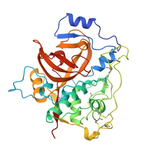
- The protein undergoes proteolytic processing from a zymogen to an active enzyme, features disulfide bonds, glycosylation, and acetylation, and its three-dimensional structure has been determined.
Experimental Guidance and Technical Tips
- The immunogen (amino acids 8-339) corresponds to the mature enzyme lacking the signal peptide, suggesting the antibody may recognize both pro- and active Cathepsin B forms. Validation in the relevant sample system is recommended.
- For Western blotting, a band near 38 kDa is expected under reducing conditions; additional bands due to processing (e.g., heavy and light chains of the mature enzyme) may be observed.
- The rabbit monoclonal format typically ensures high specificity and lot-to-lot consistency. Confirm optimal working dilutions for your specific experimental setup.
- For immunofluorescence, consider co-localization studies with lysosomal markers (e.g., LAMP1) and use appropriate fixation/permeabilization protocols.
CamelBio: Your One-Stop Sourcing Bridge
- CamelBio provides diagnostic manufacturers, laboratories, and research institutes with streamlined access to premium IVD raw materials, including validated antibody pairs, optimized monoclonal/polyclonal antibodies, bulk ancillary reagents, and rare-target sourcing. For Cathepsin B, a key enzyme in protease-related pathologies and cancer biomarker research, CamelBio offers flexible supply options to accelerate your assay development—from concept to clinic.
REQUEST A QUOTE
Our professional team will reply to you within one business day. Please feel free to contact us!