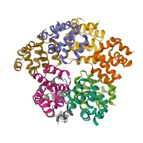
Polyclonal Antibodies
Anti-Caspase-1 Rabbit Polyclonal Antibody for WB, IF/ICC, ELISA - P29466
Item Number : CM0016000
Price varies based on specs and customizations
- Application
- WB, IF/ICC, ELISA
- Cross Reactivity
- Human, Mouse, Rat
- Protein Weight
- 45kDa

Shipping:
Contact us to get shipping details Enjoy On-time Dispatch Guarantee.
Why Choose Us
Easy ordering process, quality products, and dedicated support for your business success.
Core Product Specifications and Parameters
| Parameter | Value |
|---|---|
| Product Name | Caspase-1 Rabbit pAb |
| Remarks/Alias | ICE; P45; IL1BC; Caspase-1 |
| Species | Human |
| Gene ID | 834 |
| Immunogen | Synthetic peptide corresponding to a sequence within amino acids 201-300 of human Caspase 1 (NP_150634.1) |
| Source | Rabbit |
| Category | Polyclonal Antibodies |
| Application | WB, IF/ICC, ELISA |
| Cross Reactivity | Human, Mouse, Rat |
| SWISS | P29466 |
| Protein Weight | 45kDa |
| Shipping | Ice bag |
Biological Background: Caspase-1 Function and Localization
- Caspase-1 is a thiol protease that cleaves the precursors of interleukin-1 beta (IL1B) and interleukin 18 (IL18) into their mature active forms, playing a central role in inflammatory responses. Related references: PMID:15326478, PMID:15498465, PMID:1574116
- It also initiates pyroptosis, a lytic programmed cell death pathway, by cleaving Gasdermin-D (GSDMD), releasing its N-terminal domain to form membrane pores. Related references: PMID:26375003
- Caspase-1 activation occurs within inflammasome complexes, which assemble in response to pathogen- or damage-associated signals, leading to proximity-induced autocatalytic cleavage of the pro-enzyme.
- Alternative splicing produces multiple isoforms, including forms that lack protease activity, though their physiological roles remain incompletely characterized.
- Subcellularly, Caspase-1 is predominantly found in the cytoplasm, but a fraction associates with the cell membrane, which may facilitate its role in processing membrane-proximal substrates.
- Post-translational modifications include phosphorylation and ubiquitination, and the enzyme is initially synthesized as an inactive zymogen that requires proteolytic cleavage for activation.
- Tissue expression is highest in spleen and lung, with detection in liver, heart, small intestine, colon, thymus, prostate, skeletal muscle, peripheral blood leukocytes, kidney, and testis, but not in brain.
Experimental Guidance and Technical Tips
- The immunogen corresponds to a central region (aa 201-300) of human Caspase-1. For Western blotting, the expected molecular weight of the pro-form is around 45 kDa; upon activation, cleavage products may be observed at smaller sizes. Consider using cell lysates treated with inflammasome activators (e.g., LPS/ATP) to monitor cleavage.
- For immunofluorescence/immunocytochemistry, permeabilization is required to detect cytoplasmic and membrane-localized Caspase-1. Include appropriate controls and validate the staining pattern in the relevant cellular context.
- This antibody is predicted to cross-react with mouse and rat owing to sequence homology in the immunogen region; however, verify reactivity in your specific experimental system through preliminary tests.
CamelBio: Your One-Stop Sourcing Bridge
CamelBio connects diagnostic manufacturers and research laboratories with a comprehensive range of IVD raw materials, including validated antibodies for key targets in inflammation and cell death pathways. We offer efficient sourcing for Caspase-1 antibodies and other pyroptosis-related reagents, alongside ancillary raw materials and technical consultation to accelerate your assay development from concept to clinic. Leverage our supply network to secure bulk quantities, customized antibody pairs, and rare target reagents with streamlined logistics.
Product Datasheet
Anti-Caspase-1 Rabbit Polyclonal Antibody for WB, IF/ICC, ELISA - P29466
REQUEST A QUOTE
Our professional team will reply to you within one business day. Please feel free to contact us!