Monoclonal Antibodies
Anti-CAMK2A Rabbit Monoclonal Antibody for WB, IF-P, ELISA - Q9UQM7
Item Number : CM0006816
Price varies based on specs and customizations
- Application
- WB, IF-P, ELISA
- Cross Reactivity
- Human, Mouse, Rat
- Protein Weight
- 54kDa

Shipping:
Contact us to get shipping details Enjoy On-time Dispatch Guarantee.
Why Choose Us
Easy ordering process, quality products, and dedicated support for your business success.
Core Product Specifications and Parameters
| Parameter | Value |
|---|---|
| Product Name | CAMK2A Rabbit mAb |
| Remarks/Alias | CAMKA; MRD53; MRT63; CaMKIIalpha; CaMKIINalpha; CAMK2A |
| Species | Human |
| GeneID (Human) | 815 |
| Immunogen | Synthetic peptide within amino acids 201-300 of human CAMK2A (NP_741960.1) |
| Source | Rabbit |
| Category | Monoclonal Antibodies |
| Application | WB, IF-P, ELISA |
| Cross Reactivity | Human, Mouse, Rat |
| SWISS | Q9UQM7 |
| Protein Weight | 54kDa |
| Shipping | Ice bag |
Biological Background: CAMK2A Function and Localization
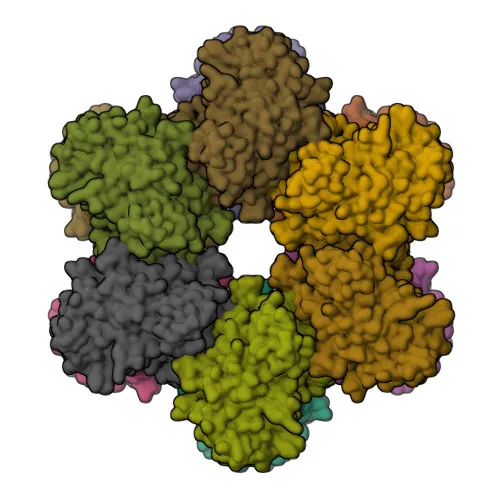
- Calcium/calmodulin-dependent protein kinase type II subunit alpha (CAMK2A) is a serine/threonine-protein kinase that becomes autonomously active upon Ca²⁺/calmodulin binding and autophosphorylation, playing a central role in synaptic plasticity, neurotransmitter release, and long-term potentiation (LTP).
Related references: PMID:14722083 - CAMK2A is a key component of the NMDAR signaling complex at excitatory synapses, where it regulates AMPA receptor (AMPAR) trafficking and synaptic strength, thus modulating excitatory synaptic transmission (By similarity).
- Beyond synaptic functions, CAMK2A phosphorylates multiple substrates including FOXO3 to promote its transcriptional activity (PubMed:23805378) and ETS1 to reduce its DNA-binding affinity in response to calcium signaling (By similarity).
Related references: PMID:23805378 - In the immune context, CAMK2A mediates interferon signaling by phosphorylating STAT1 upon IFN-gamma stimulation (PubMed:11972023) and IFN-beta stimulation (PubMed:35568036). It also phosphorylates PSAT1 to inhibit ferroptosis via GPX4 stabilization (PubMed:40281343).
Related references: PMID:11972023, PMID:35568036, PMID:40281343 - Subcellularly, CAMK2A localizes to the postsynaptic density, dendritic spines, and dendrites, consistent with its role in synaptic function and neuronal morphology regulation.
- CAMK2A is subject to alternative splicing and post-translational modifications, including palmitoylation and autophosphorylation, which modulate its activity and localization (UniProt keywords).
- Mutations in CAMK2A are associated with intellectual disability and neurodevelopmental disorders, highlighting its essential role in brain development and function.
Experimental Guidance and Technical Tips
- The immunogen corresponds to a region within amino acids 201-300 of human CAMK2A, a sequence conserved in mouse and rat, supporting the observed cross-reactivity.
- For Western blotting, a band at approximately 54 kDa is expected; consider using neuronal tissue lysates (e.g., brain cortex) or cell lines expressing CAMK2A as positive controls.
- In immunofluorescence-paraffin (IF-P) experiments, target localization to dendritic spines and postsynaptic densities in brain sections; optimal fixation and permeabilization conditions should be validated.
- For ELISA applications, a synthetic peptide or recombinant protein spanning the immunogen region may serve as coating antigen; assay conditions require individual optimization.
CamelBio: Your One-Stop Sourcing Bridge
CamelBio offers a comprehensive portfolio of IVD raw materials, including validated monoclonal antibodies like anti-CAMK2A, for neuroscience and signal transduction research. As a one-stop sourcing partner for diagnostic manufacturers and laboratories, we provide custom antibody pairs, bulk reagents, and expert support to accelerate your development pipeline from concept to clinic.
REQUEST A QUOTE
Our professional team will reply to you within one business day. Please feel free to contact us!