Polyclonal Antibodies
Anti-CACNA1H Rabbit Polyclonal Antibody for IF-P, ELISA - O95180
Item Number : CM0022944
Price varies based on specs and customizations
- Application
- IF-P, ELISA
- Cross Reactivity
- Rat
- Protein Weight
- 259kDa

Shipping:
Contact us to get shipping details Enjoy On-time Dispatch Guarantee.
Why Choose Us
Easy ordering process, quality products, and dedicated support for your business success.
Core Product Specifications and Parameters
| Parameter | Value |
|---|---|
| Product Name | CACNA1H Rabbit pAb |
| Remarks/Alias | ECA6; EIG6; HALD4; Cav3.2; CACNA1HB; CACNA1H |
| Species | Human |
| Gene ID (Human) | 8912 |
| Gene ID | 8912 |
| Immunogen | A synthetic peptide corresponding to a sequence within amino acids 2250-2353 of human CACNA1H (NP_066921.2). |
| Source | Rabbit |
| Category | Polyclonal Antibodies |
| Application | IF-P, ELISA |
| Cross Reactivity | Rat |
| SWISS | O95180 |
| Protein Weight | 259kDa |
| Shipping | Ice bag |
Biological Background: CACNA1H Function and Localization
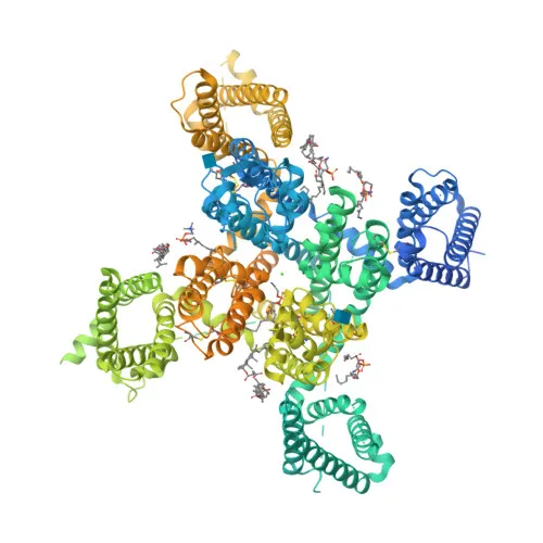
- CACNA1H encodes the alpha-1H subunit of the voltage-dependent T-type calcium channel, also known as Cav3.2, a member of the low-voltage-activated (LVA) calcium channel family.
- The gene undergoes alternative splicing, producing multiple isoforms with potentially distinct functional and tissue distributions.
- Voltage-sensitive calcium channel that mediates T-type calcium currents, opening at relatively negative membrane potentials and inactivating in a voltage-dependent manner. Related references: PMID:27149520, PMID:9670923, PMID:9930755
- T-type channels serve pacemaking functions in central neurons and cardiac nodal cells, and support calcium signaling in secretory cells and vascular smooth muscle.
- May modulate firing patterns of neurons, influencing neuronal excitability. Related references: PMID:15048902
- In the adrenal zona glomerulosa, participates in the signaling pathway leading to aldosterone production in response to angiotensin II or hyperkalemia. Related references: PMID:25907736, PMID:27729216
- Tissue expression: Detected at protein level in adrenal zona glomerulosa. Highly expressed in kidney, liver, heart, and brain regions (amygdala, caudate nucleus, putamen). Also expressed in testis germ cells. Related references: PMID:25907736, PMID:27729216, PMID:9670923, PMID:9930755, PMID:11751928
- The channel is a glycoprotein and binds calcium and zinc ions; it localizes to the cell membrane as a multi-pass transmembrane protein.
- Disease-associated variants have been linked to epilepsy, underscoring its role in neuronal excitability disorders.
Experimental Guidance and Technical Tips
- Given the immunogen is a synthetic peptide from the C-terminal region (aa 2250-2353), consider verifying specificity by peptide blocking experiments.
- For immunofluorescence (IF-P), optimize fixation and permeabilization conditions for membrane proteins; antigen retrieval may be required for paraffin sections.
- This antibody cross-reacts with rat CACNA1H, making it suitable for rodent model studies; validate in the relevant tissue system.
- In ELISA applications, the immunizing peptide can serve as a positive control; include appropriate secondary antibody controls.
- Since CACNA1H is a large multi-pass membrane protein, consider using mild detergents for protein extraction to preserve epitope accessibility.
CamelBio: Your One-Stop Sourcing Bridge
CamelBio offers reliable sourcing for IVD raw materials, including this anti-CACNA1H polyclonal antibody, to support your ion channel and neuroscience research. As a one-stop bridge, we provide validated antibody pairs, bulk ancillary reagents, and customized procurement for rare targets, streamlining your development from concept to clinic. Whether you need monoclonal alternatives or complementary detection reagents, CamelBio ensures consistent supply to meet your manufacturing and research demands.
REQUEST A QUOTE
Our professional team will reply to you within one business day. Please feel free to contact us!