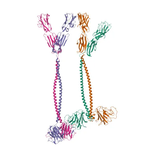
Monoclonal Antibodies
Anti-BTN3A1/2/3 Monoclonal Antibody for FC - O00481 / P78410 / O00478
Item Number : CM0010074
Price varies based on specs and customizations
- Application
- FC
- Cross Reactivity
- Human
- Protein Weight
- 58kDa

Shipping:
Contact us to get shipping details Enjoy On-time Dispatch Guarantee.
Why Choose Us
Easy ordering process, quality products, and dedicated support for your business success.
Core Product Specifications and Parameters
| Parameter | Value |
|---|---|
| Product Name | Rabbit anti-Human BTN3A1/2/3 mAb |
| Remarks/Alias | BTF5; BT3.1; CD277; BTN3.1 |
| Species | Human |
| GeneID (Human) | 11119/11118/10384 |
| GeneID | 11119/11118/10384 |
| Immunogen | Recombinant protein, Recombinant fusion protein containing a sequence corresponding to amino acids 470-630 of human BTN3A1 (NP_008979.3) |
| Source | Rabbit |
| Category | Monoclonal Antibodies |
| Application | FC |
| Cross Reactivity | Human |
| SWISS | O00481/P78410/O00478 |
| Protein Weight | 58kDa |
| Shipping | Ice bag |
Biological Background: BTN3A1 Function and Localization
- BTN3A1 (Butyrophilin subfamily 3 member A1), also known as BTF5, BT3.1, or CD277, is a transmembrane glycoprotein belonging to the butyrophilin family, characterized by immunoglobulin-like domains.
- It plays a critical role in T-cell activation and adaptive immunity by regulating the proliferation of activated T cells and the release of cytokines such as IFNG.
- BTN3A1 mediates the response of T cells toward infected and transformed cells that exhibit elevated levels of phosphorylated metabolites (e.g., isopentenyl pyrophosphate), acting as a sensor for metabolic dysregulation.
- The protein localizes to the cell membrane, consistent with its function in immune surveillance and cell–cell communication.
- BTN3A1 is widely expressed; at the protein level, it is detected on T cells, natural killer cells, dendritic cells, and macrophages, with high transcript levels in heart, pancreas, and lung.
- Post-translational modifications include disulfide bond formation (essential for immunoglobulin domain stability) and glycosylation, which may influence protein folding and ligand interaction.
- Alternative splicing generates multiple isoforms, expanding functional diversity; the canonical sequence includes a signal peptide, extracellular immunoglobulin domains, a transmembrane helix, and a cytoplasmic tail.
Experimental Guidance and Technical Tips
- The immunogen corresponds to the C-terminal region (amino acids 470–630) of human BTN3A1, a domain that may include part of the cytoplasmic tail; this design likely contributes to the antibody’s specificity for BTN3A1 and possibly cross-reactivity with BTN3A2/A3.
- This antibody is directly conjugated to , a dye optimized for flow cytometry; no secondary antibody is required. Consider titrating the antibody to determine the optimal concentration for your specific cell type and staining conditions.
- For FC experiments, ensure appropriate single-stain controls and compensation when using multicolor panels. Dead cell exclusion and Fc receptor blocking may improve signal-to-noise ratio.
- The product is recommended for human samples; cross-reactivity with other species has not been reported. Validate performance in your target cell population (e.g., T cells, NK cells) using positive and negative controls.
CamelBio: Your One-Stop Sourcing Bridge
CamelBio serves diagnostic manufacturers, labs, and research institutes as a comprehensive IVD raw-material sourcing partner, providing access to validated antibody pairs, optimized monoclonal/polyclonal antibodies, bulk ancillary reagents, and rare target raw materials. This BTN3A1/2/3 monoclonal antibody supports immune function research and T-cell studies, aligning with our portfolio of high-quality immunological tools. For your full workflow needs—from concept to clinic—CamelBio delivers reliable, scalable solutions.
Product Datasheet
Anti-BTN3A1/2/3 Monoclonal Antibody for FC - O00481 / P78410 / O00478
REQUEST A QUOTE
Our professional team will reply to you within one business day. Please feel free to contact us!