Monoclonal Antibodies
Anti-BST2 Rabbit Monoclonal Antibody for WB, IHC-P, IF/ICC, ELISA - Q10589
Item Number : CM0007383
Price varies based on specs and customizations
- Application
- WB, IHC-P, IF/ICC, ELISA
- Cross Reactivity
- Human
- Protein Weight
- 20kDa

Shipping:
Contact us to get shipping details Enjoy On-time Dispatch Guarantee.
Why Choose Us
Easy ordering process, quality products, and dedicated support for your business success.
Core Product Specifications and Parameters
| Parameter | Value |
|---|---|
| Product Name | BST2 Rabbit mAb |
| Remarks/Alias | CD317; HM1.24; TETHERIN; BST2 |
| Species | Human |
| Gene ID (Human) | 684 |
| Gene ID | 684 |
| Immunogen | Recombinant fusion protein containing a sequence corresponding to amino acids 49-160 of human BST2 (NP_004326.1) |
| Source | Rabbit |
| Category | Monoclonal Antibodies |
| Application | WB, IHC-P, IF/ICC, ELISA |
| Cross Reactivity | Human |
| SWISS | Q10589 |
| Protein Weight | 20kDa |
| Shipping | Ice bag |
Biological Background: BST2 Function and Localization
- BST2 (bone marrow stromal antigen 2), also known as Tetherin, CD317, or HM1.24 antigen, is an interferon-induced host restriction factor with two major isoforms that differ in antiviral activity and susceptibility to viral countermeasures. Isoform 2 is notably resistant to HIV-1 Vpu-mediated degradation.
Related references: PMID:23028328 PMID:26172439 - The primary antiviral mechanism involves direct physical tethering of nascent enveloped virions to the cell membrane, preventing their release. Tethered virions can be internalized via endocytosis and degraded, thereby restricting cell-free viral spread.
Related references: PMID:18200009 PMID:18342597 PMID:19036818 - BST2 restricts a broad range of enveloped viruses, including retroviruses (HIV-1, HIV-2, SIV), filoviruses (Ebola, Marburg), flaviviruses (HCV), herpesviruses (KSHV), coronaviruses (SARS-CoV), and many others.
Related references: PMID:18200009 PMID:18342597 PMID:19179289 - Beyond viral restriction, BST2 can inhibit cell surface proteolytic activity of MMP14, leading to reduced MMP15 activation and inhibition of cell growth and migration.
Related references: PMID:22065321 - In plasmacytoid dendritic cells, BST2 stimulates LILRA4/ILT7 signaling, providing negative feedback to IFN production during viral infection.
Related references: PMID:19564354 PMID:26172439 - Subcellular localization includes the Golgi apparatus (trans-Golgi network), cell membrane, membrane rafts, cytoplasm, apical cell membrane, and late endosomes, consistent with its role in viral trafficking and immune signaling.
- BST2 is predominantly expressed in liver, lung, heart, and placenta, with lower levels in pancreas, kidney, skeletal muscle, and brain. At the protein level, it is highly expressed on B-cells, T-cells, monocytes, NK cells, and dendritic cells, and is overexpressed in multiple myeloma cells.
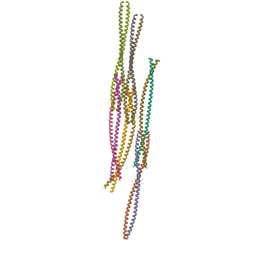
- Structurally, BST2 features a signal-anchor, single-pass transmembrane domain, coiled-coil ectodomain, C-terminal GPI-anchor, and disulfide bonds, which are critical for its tethering function and dimerization.
Experimental Guidance and Technical Tips
- The immunogen covers amino acids 49–160 of human BST2, a region within the N-terminal ectodomain. This region is likely well-exposed on the cell surface, making the antibody suitable for detecting membrane-bound BST2 in applications such as IF/ICC and IHC-P.
- For Western blotting, the predicted molecular weight of BST2 is approximately 20 kDa under reducing conditions; however, glycosylation and dimerization may lead to higher molecular weight species. Optimization of reducing conditions and loading controls is recommended.
- In immunohistochemistry and immunofluorescence, verify BST2 expression in positive control tissues such as liver, lung, or multiple myeloma cells. Membrane and intracellular staining should be anticipated given its Golgi and endosomal localization.
- This antibody is validated for human samples only; cross-reactivity with other species has not been determined. For ELISA, consider pairing with a matched detection antibody for sensitive antigen capture.
- Since BST2 is an IFN-inducible gene, treatment of cells with interferon may upregulate target expression and enhance detection sensitivity.
CamelBio: Your One-Stop Sourcing Bridge
CamelBio serves as a one-stop sourcing platform for IVD raw materials, providing diagnostic manufacturers and researchers with validated antibodies like this anti-BST2 monoclonal antibody. Our supply capabilities extend to antibody pairs, recombinant proteins, and ancillary reagents for immunodiagnostic development. Whether you are investigating antiviral restrictions or targeting multiple myeloma biomarkers, CamelBio can support your project with rare-target material sourcing and expert technical consultation.
Product Datasheet
Anti-BST2 Rabbit Monoclonal Antibody for WB, IHC-P, IF/ICC, ELISA - Q10589
REQUEST A QUOTE
Our professional team will reply to you within one business day. Please feel free to contact us!