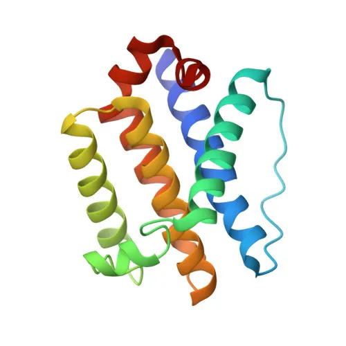
Polyclonal Antibodies
Anti-BOK Rabbit Polyclonal Antibody for WB, IF/ICC, IHC-P, ELISA - Q9UMX3
Item Number : CM0020643
Price varies based on specs and customizations
- Application
- WB, IF/ICC, IHC-P, ELISA
- Cross Reactivity
- Human, Mouse, Rat
- Protein Weight
- 23 kDa

Shipping:
Contact us to get shipping details Enjoy On-time Dispatch Guarantee.
Why Choose Us
Easy ordering process, quality products, and dedicated support for your business success.
Core Product Specifications and Parameters
| Parameter | Value |
|---|---|
| Product Name | BOK Rabbit pAb |
| Remarks/Alias | BOKL; BCL2L9; BOK |
| Species | Human |
| Gene ID (Human) | 666 |
| Gene ID | 666 |
| Immunogen | Synthetic Peptide|A synthetic peptide corresponding to a sequence within amino acids 1-100 of human BOK (NP_115904.1). |
| Source | Rabbit |
| Category | Polyclonal Antibodies |
| Application | WB, IF/ICC, IHC-P, ELISA |
| Cross Reactivity | Human, Mouse, Rat |
| SWISS | Q9UMX3 |
| Protein Weight | 23 kDa |
| Shipping | Ice bag |
Biological Background: BOK Function and Localization
- BOK (Bcl-2-related ovarian killer protein), also known as Bcl-2-like protein 9, is a pro-apoptotic member of the BCL-2 family that promotes intrinsic apoptosis through both BAX/BAK1-dependent and -independent mechanisms. Related references: PMID:15102863 PMID:27076518
- Upon endoplasmic reticulum (ER) stress, BOK mediates mitochondrial apoptosis by activating BAX/BAK1 and positively regulating PERK-dependent unfolded protein response.
- In the DNA damage response, BOK triggers intrinsic apoptosis in a TP53-dependent manner. Related references: PMID:15102863
- BOK contributes to granulosa cell apoptosis through CASP3 activation, highlighting a role in ovarian follicle atresia. Related references: PMID:20673843
- Beyond classical apoptosis, BOK can induce autophagy by disrupting the MCL1-BECN1 interaction and has been implicated in regulating trophoblast cell proliferation during placental development. Related references: PMID:24113155 PMID:19942931
- In neuronal contexts, BOK exhibits an anti-apoptotic function by regulating calcium homeostasis and mitochondrial bioenergetics, thereby inhibiting poly(ADP-ribose) polymerase-dependent cell death.
- Subcellular localization includes mitochondrion (inner and outer membranes), endoplasmic reticulum, Golgi apparatus, endosomes, nucleus, and cytoplasm, consistent with its multifaceted roles in cell death and survival.
- Tissue specificity: Highly expressed in oocytes and granulosa cells of ovarian follicles, with strongest expression in primordial/primary follicle granulosa cells.
Experimental Guidance and Technical Tips
- For western blotting, a protein band around 23 kDa may be expected. Use positive control lysates from human, mouse, or rat tissues based on the stated cross-reactivity.
- In immunocytochemistry and immunohistochemistry, verify staining patterns against the known subcellular distribution of BOK (mitochondria, ER, Golgi, endosomes, nucleus).
- The immunogen corresponds to amino acids 1–100 of human BOK (NP_115904.1). Consider epitope accessibility in your sample preparation and assay conditions.
- For ELISA applications, the antibody may serve as a capture or detection reagent. Optimization of coating concentrations and detection systems is recommended.
CamelBio: Your One-Stop Sourcing Bridge
CamelBio provides a comprehensive sourcing solution for IVD raw materials, from validated antibody pairs to optimized polyclonal antibodies and bulk ancillary reagents. This anti-BOK antibody supports research in apoptosis and cancer biology, and our team can assist with sourcing rare targets, custom development, and technical consultation to accelerate your diagnostic and research projects from concept to clinic.
Product Datasheet
Anti-BOK Rabbit Polyclonal Antibody for WB, IF/ICC, IHC-P, ELISA - Q9UMX3
REQUEST A QUOTE
Our professional team will reply to you within one business day. Please feel free to contact us!