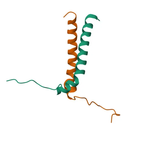
Polyclonal Antibodies
Anti-BNIP3 Polyclonal Antibody for WB, IHC-P, ELISA - Q12983
Item Number : CM0030765
Price varies based on specs and customizations
- Application
- WB, IHC-P, ELISA
- Cross Reactivity
- Human, Mouse, Rat
- Protein Weight
- 22kDa

Shipping:
Contact us to get shipping details Enjoy On-time Dispatch Guarantee.
Why Choose Us
Easy ordering process, quality products, and dedicated support for your business success.
Core Product Specifications and Parameters
| Parameter | Value |
|---|---|
| Product Name | BNIP3 Rabbit pAb |
| Remarks/Alias | NIP3; HABON; BNIP3 |
| Species | Human |
| GeneID (human) | 664 |
| GeneID | 664 |
| Immunogen | Recombinant fusion protein containing a sequence corresponding to amino acids 40-120 of human BNIP3 (NP_004043.3) |
| Source | Rabbit |
| Category | Polyclonal Antibodies |
| Application | WB, IHC-P, ELISA |
| Cross Reactivity | Human, Mouse, Rat |
| SWISS | Q12983 |
| Protein Weight | 22kDa |
| Shipping | Ice bag |
Biological Background: BNIP3 Function and Localization
- BNIP3 (BCL2/adenovirus E1B 19 kDa protein-interacting protein 3) is a pro-apoptotic member of the BCL2 family that can overcome BCL2-mediated survival suppression and participate in repartitioning calcium between intracellular stores.
- It plays a key role in mitochondrial quality control by interacting with SPATA18/MIEAP, driving the degradation of damaged mitochondrial proteins through the MALM (mitochondrial protein catabolic process) pathway.
- Together with BNIP3L/NIX, BNIP3 regulates the formation of a pore in the mitochondrial double membrane, enabling the translocation of lysosomal proteins into the mitochondrial matrix for organelle turnover.
- BNIP3 is involved in calprotectin (S100A8/A9)-induced cell death, linking innate immune signaling to mitochondrial dysfunction and apoptotic execution.
- The protein localizes to the mitochondrial outer membrane via a C-terminal transmembrane domain, and its activity is modulated by phosphorylation.
- BNIP3 has been implicated in host-virus interactions, counteracting viral anti-apoptotic factors such as adenovirus E1B 19K, and is part of the reference proteome with a calculated molecular weight of ~21.5 kDa.
Experimental Guidance and Technical Tips
- For Western blot, the antibody detects a band at approximately 22 kDa under reducing conditions; consider using a 12–15% SDS-PAGE gel for optimal resolution.
- Validate reactivity in human, mouse, or rat cell lines or tissue lysates; include appropriate positive (e.g., cells treated with hypoxia or apoptosis inducers) and negative controls.
- For IHC-P, perform antigen retrieval (e.g., citrate buffer, pH 6.0) and titrate the primary antibody to minimize background; validate in relevant tissue sections (e.g., heart, skeletal muscle, or tumor biopsies).
- In ELISA, consider using the antibody as a capture or detection reagent after confirming binding affinity against recombinant or native BNIP3.
- Since BNIP3 is a membrane protein, use mild lysis buffers (e.g., RIPA with protease inhibitors) and avoid boiling samples excessively to prevent aggregation.
CamelBio: Your One-Stop Sourcing Bridge
CamelBio provides diagnostic manufacturers and researchers with a comprehensive one-stop access to IVD raw materials, including validated antibodies and ancillary reagents for mitochondrial biology and cell death research. This BNIP3 polyclonal antibody supports your R&D and assay development programs, and we can further supply optimized monoclonal/polyclonal antibody pairs, bulk ancillary reagents, and rare target raw-material sourcing to accelerate your project from concept to clinic.
REQUEST A QUOTE
Our professional team will reply to you within one business day. Please feel free to contact us!