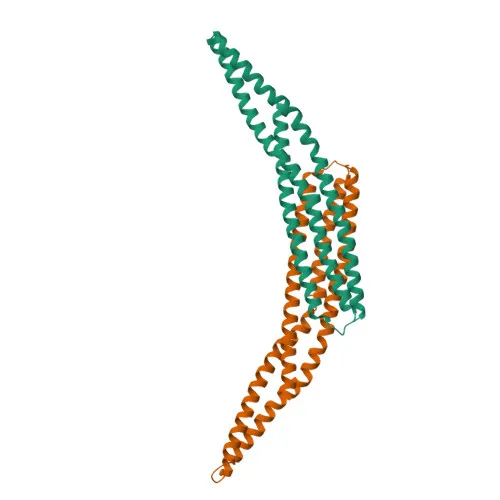
Monoclonal Antibodies
Anti-BIN1 Monoclonal Antibody for WB, IHC-P, ELISA - O00499
Item Number : CM0003542
Price varies based on specs and customizations
- Application
- WB, IHC-P, ELISA
- Cross Reactivity
- Human, Mouse, Rat
- Protein Weight
- 65kDa

Shipping:
Contact us to get shipping details Enjoy On-time Dispatch Guarantee.
Why Choose Us
Easy ordering process, quality products, and dedicated support for your business success.
Core Product Specifications and Parameters
| Parameter | Value |
|---|---|
| Product Name | BIN1 Rabbit mAb |
| Remarks/Alias | CNM2; AMPH2; AMPHL; SH3P9; BIN1 |
| Species | Human |
| Gene ID (Human) | 274 |
| Gene ID | 274 |
| Immunogen | Recombinant fusion protein containing a sequence corresponding to amino acids 400-593 of human BIN1 (NP_647601.1) |
| Source | Rabbit |
| Category | Monoclonal Antibodies |
| Application | WB, IHC-P, ELISA |
| Cross Reactivity | Human, Mouse, Rat |
| SWISS | O00499 |
| Protein Weight | 65kDa |
| Shipping | Ice bag |
Biological Background: BIN1 Function and Localization
- BIN1 (Myc box-dependent-interacting protein 1), also known as Amphiphysin II, is a key regulator of plasma membrane curvature and remodeling, essential for T-tubule formation in muscle cells. Related references: PMID:24755653
- It acts as a negative regulator of endocytosis and is involved in intracellular vesicle sorting and BACE1 trafficking, influencing amyloid-beta production. Related references: PMID:27179792
- BIN1 may regulate endocytosis in neuronal circuits, affecting the internalization of PHF-tau aggregates.
- It is involved in regulating MYC activity and cell proliferation. Related references: PMID:8782822
- BIN1 possesses actin bundling activity and stabilizes actin filaments against depolymerization in vitro. Related references: PMID:28893863
- Subcellularly, BIN1 localizes to the nucleus, cytoplasm, endosomes, cell membrane, sarcolemma, and T-tubules.
- Tissue expression is ubiquitous, with highest levels in brain and muscle; isoform IIA is brain-specific, and the major isoform is highly expressed in skeletal muscle.
- Key features include alternative splicing, a coiled-coil domain, an SH3 domain, and roles in development, differentiation, and host-virus interactions.
Experimental Guidance and Technical Tips
- The immunogen corresponds to amino acids 400–593, the C-terminal region containing the SH3 domain. This design targets a functional domain, potentially recognizing multiple isoforms.
- For Western blot, BIN1 is predicted at ~65 kDa; validate the observed band size in your sample lysates, considering possible posttranslational modifications or isoform variants.
- For IHC-P, consider optimizing antigen retrieval and antibody dilution using tissues with known high expression (e.g., brain, skeletal muscle).
- For ELISA, use recombinant BIN1 protein as a standard to assess binding affinity and specificity.
- Cross-reactivity is reported for human, mouse, and rat; verify reactivity in other species if needed.
- This antibody is provided in a format suitable for routine applications; for sandwich immunoassays, evaluate as a capture or detection antibody in combination with a suitable pair.
CamelBio: Your One-Stop Sourcing Bridge
CamelBio supports diagnostic manufacturers, laboratories, and research institutes with a comprehensive range of IVD raw materials, technical services, and consulting—from concept to clinic. Our portfolio includes validated monoclonal antibodies, recombinant proteins, and ancillary reagents for membrane trafficking and neurodegenerative disease research. For BIN1-related projects, we can help source rare targets, optimized antibody pairs, and bulk reagents to accelerate your assay development and scale-up.
REQUEST A QUOTE
Our professional team will reply to you within one business day. Please feel free to contact us!