Polyclonal Antibodies
Anti-BEST1 Rabbit Polyclonal Antibody for WB, ELISA - O76090
Item Number : CM0030348
Price varies based on specs and customizations
- Application
- WB, ELISA
- Cross Reactivity
- Human, Mouse, Rat
- Protein Weight
- 68kDa

Shipping:
Contact us to get shipping details Enjoy On-time Dispatch Guarantee.
Why Choose Us
Easy ordering process, quality products, and dedicated support for your business success.
Core Product Specifications and Parameters
| Parameter | Value |
|---|---|
| Product Name | BEST1 Rabbit pAb |
| Remarks/Alias | ARB; BMD; BEST; RP50; VMD2; TU15B; Best1V1Delta2; BEST1 |
| Species | Human |
| GeneID (Human) | 7439 |
| GeneID | 7439 |
| Immunogen | A synthetic peptide corresponding to a sequence within amino acids 171-270 of human BEST1 (NP_004174.1). |
| Source | Rabbit |
| Category | Polyclonal Antibodies |
| Application | WB, ELISA |
| Cross Reactivity | Human, Mouse, Rat |
| SWISS | O76090 |
| Protein Weight | 68kDa |
| Shipping | Ice bag |
Biological Background: Bestrophin-1 Function and Localization
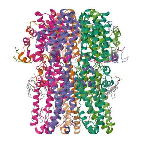
- Bestrophin-1 (BEST1), also designated TU15B or vitelliform macular dystrophy protein 2, is a member of the bestrophin family encoded by the BEST1 gene.
- It operates as a calcium-activated ligand-gated anion channel that conducts chloride and bicarbonate ions (PubMed:11904445, PubMed:12907679, PubMed:18179881). A partially open conformation limits chloride flux (PubMed:35789156). Related references: PMID:11904445 PMID:12907679 PMID:18179881 PMID:35789156
- In astrocytes, BEST1 facilitates Ca²⁺-dependent release of glutamate upon PAR-1 activation, influencing synaptic plasticity (By similarity).
- It also transports D-serine and glutamate via norepinephrine signaling, modulating heterosynaptic long-term depression and metaplasticity (By similarity).
- Additionally, BEST1 participates in tonic GABA release from cerebellar glial cells (By similarity).
- Subcellularly, BEST1 localizes to the basolateral cell membrane and is predominantly expressed in the retinal pigment epithelium, where it plays a vital role in retinal health.
- The protein contains multiple transmembrane domains, undergoes phosphorylation, and is associated with macular dystrophies such as Best vitelliform macular dystrophy.
Experimental Guidance and Technical Tips
- The immunogen corresponds to a cytoplasmic region (amino acids 171–270) of human BEST1; antibodies raised against linear epitopes may perform well in denaturing Western blot. Consider optimizing blocking and antibody dilution for your specific sample.
- For ELISA, coat with synthetic peptide or recombinant protein fragment; include appropriate positive and negative controls to assess specificity.
- With cross-reactivity to mouse and rat, this antibody is suitable for rodent retinal and brain tissue studies. Validate in the relevant species and tissue type to confirm performance.
- The predicted molecular weight is 68 kDa; in WB, confirm the band appearance and any possible post-translational modifications or degradation products.
CamelBio: Your One-Stop Sourcing Bridge
CamelBio provides diagnostic manufacturers, labs, and research institutes with one‑stop access to IVD raw materials, technical services, and consulting. For retinal biology and anion channel studies, we offer validated antibody pairs, optimized monoclonal/polyclonal antibodies, and bulk ancillary reagents. Our sourcing solutions cover every stage from concept to clinic, accelerating your assay development.
REQUEST A QUOTE
Our professional team will reply to you within one business day. Please feel free to contact us!