Polyclonal Antibodies
Anti-Bak Rabbit Polyclonal Antibody for WB, ELISA - Q16611
Item Number : CM0023664
Price varies based on specs and customizations
- Application
- WB, ELISA
- Cross Reactivity
- Human, Mouse
- Protein Weight
- 23kDa

Shipping:
Contact us to get shipping details Enjoy On-time Dispatch Guarantee.
Why Choose Us
Easy ordering process, quality products, and dedicated support for your business success.
Core Product Specifications and Parameters
| Parameter | Value |
|---|---|
| Product Name | Bak Rabbit pAb |
| Remarks/Alias | BAK; CDN1; BCL2L7; BAK-LIKE; Bak |
| Species | Human |
| Gene ID (Human) | 578 |
| Gene ID | 578 |
| Immunogen | A synthetic peptide corresponding to a sequence within amino acids 1-100 of human Bak (NP_001179.1) |
| Source | Rabbit |
| Category | Polyclonal Antibodies |
| Application | WB, ELISA |
| Cross Reactivity | Human, Mouse |
| SWISS | Q16611 |
| Protein Weight | 23kDa |
| Shipping | Ice bag |
Biological Background: Bak Function and Localization
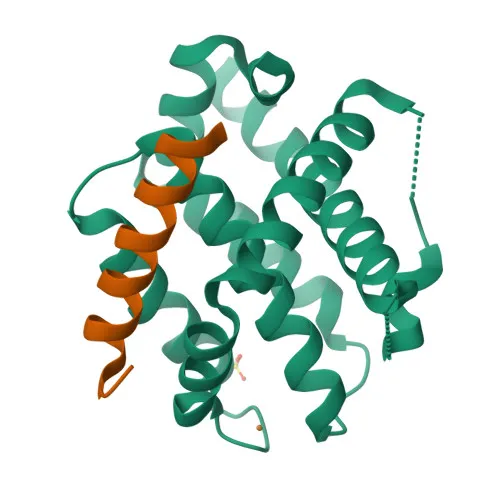
- Bak (Bcl-2 homologous antagonist/killer), also known as apoptosis regulator BAK or Bcl2-L-7, is a pro-apoptotic member of the Bcl-2 protein family.
- Encoded by the BAK1 gene (Gene ID: 578), it plays a critical role in the intrinsic mitochondrial apoptotic pathway.
- Upon apoptotic stimuli, Bak oligomerizes and forms pores in the mitochondrial outer membrane (MOM), leading to permeabilization and release of apoptogenic factors such as cytochrome c into the cytosol.
- This release triggers the activation of caspase 9 and downstream effector caspases, executing the cell death program.
- Bak is primarily localized to the mitochondrial outer membrane, consistent with its function in apoptosis regulation.
- It is widely expressed across human tissues, with the highest expression levels in the heart and skeletal muscle.
- Post-translational modifications, including acetylation and zinc binding, may regulate Bak activity, and alternative splicing generates multiple isoforms.
- The protein interacts with host factors during viral infection, highlighting its role in host-virus interactions.
Experimental Guidance and Technical Tips
- The antibody was raised against a synthetic peptide corresponding to the N-terminal 1–100 amino acids of human Bak; it is expected to recognize full-length endogenous Bak protein.
- For Western blotting, the predicted molecular weight is ~23 kDa; consider using mitochondrial-enriched fractions or positive control lysates from heart or skeletal muscle where Bak expression is highest.
- When performing ELISA, titrate the antibody to determine optimal coating and detection concentrations, and validate specificity using recombinant Bak protein or peptide competition assays.
- Cross-reactivity with mouse is reported; however, validate in your specific sample system, as detection may vary with sample preparation and epitope accessibility.
- Since Bak is a mitochondrial membrane protein, ensure proper lysis buffers and denaturing conditions to expose the epitope.
CamelBio: Your One-Stop Sourcing Bridge
CamelBio offers diagnostic manufacturers and researchers convenient access to high-quality IVD raw materials like this anti-Bak antibody, supporting apoptosis and cancer research. We supply validated antibody pairs, optimized polyclonal and monoclonal antibodies, and ancillary reagents in bulk, along with rare-target raw-material sourcing to accelerate your assay development from concept to clinic.
REQUEST A QUOTE
Our professional team will reply to you within one business day. Please feel free to contact us!