Monoclonal Antibodies
Anti-Bak Monoclonal Antibody for WB, IHC-P, ELISA - Q16611
Item Number : CM0006172
Price varies based on specs and customizations
- Application
- WB, IHC-P, ELISA
- Cross Reactivity
- Human, Monkey
- Protein Weight
- 23kDa

Shipping:
Contact us to get shipping details Enjoy On-time Dispatch Guarantee.
Why Choose Us
Easy ordering process, quality products, and dedicated support for your business success.
Core Product Specifications and Parameters
| Parameter | Value |
|---|---|
| Product Name | Bak Rabbit mAb |
| Remarks/Alias | BAK; CDN1; BCL2L7; BAK-LIKE; Bak |
| Species | Human |
| GeneID (Human) | 578 |
| GeneID | 578 |
| Immunogen | Recombinant fusion protein containing a sequence corresponding to amino acids 11-82 of human Bak (NP_001179.1). |
| Source | Rabbit |
| Category | Monoclonal Antibodies |
| Application | WB, IHC-P, ELISA |
| Cross Reactivity | Human, Monkey |
| SWISS | Q16611 |
| Protein Weight | 23kDa |
| Shipping | Ice bag |
Biological Background: Bak Function and Localization
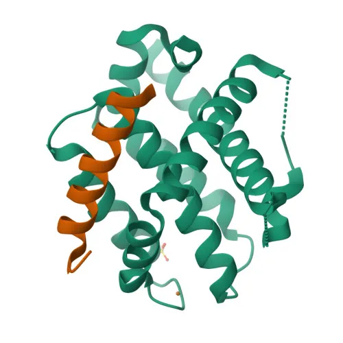
- Bak, encoded by the BAK1 gene (also known as BCL2L7, CDN1), is a pro-apoptotic member of the Bcl-2 protein family, sharing conserved BH (Bcl-2 homology) domains essential for its dimerization and function.
- As a critical effector of the mitochondrial (intrinsic) apoptotic pathway, Bak oligomerizes upon activation to form pores in the mitochondrial outer membrane (MOM), leading to MOMP (mitochondrial outer membrane permeabilization) and release of cytochrome c and other apoptogenic factors, which activates downstream caspases.
- Together with its close homolog Bax, Bak constitutes the core pore-forming machinery required for mitochondrial apoptosis, balancing cell survival signals with cell death execution.
- Ubiquitously expressed, with highest levels detected in heart and skeletal muscle, Bak is primarily localized to the mitochondrial outer membrane, tethered via its C-terminal transmembrane domain.
- Functionally, Bak is regulated by interactions with anti-apoptotic Bcl-2 members (e.g., Bcl-2, Bcl-XL) and BH3-only proteins, and its activity is modulated by post-translational modifications including acetylation and metal-ion interactions.
- Recent structural studies have elucidated the conformational changes underlying Bak activation and pore formation, highlighting its role in response to cellular stress, DNA damage, and developmental cues.
Experimental Guidance and Technical Tips
- The recombinant protein immunogen spans amino acids 11-82 of human Bak, covering a region conserved in monkeys, supporting cross-reactivity in both species; consider validating detection in your specific sample matrix.
- This monoclonal antibody is validated for Western blot (WB), immunohistochemistry-paraffin (IHC-P), and ELISA applications by the supplier; for each application, optimal dilution and protocol should be empirically determined using positive and negative controls.
- Due to the predicted molecular weight of 23 kDa (UniProt: 23.4 kDa), ensure proper separation on SDS-PAGE gels and include a suitable molecular weight marker.
CamelBio: Your One-Stop Sourcing Bridge
CamelBio serves as a reliable one-stop sourcing partner for IVD raw materials, offering a comprehensive portfolio of antibodies, antigens, and ancillary reagents to support diagnostic assay development from concept to clinic. With the anti-Bak monoclonal antibody, researchers and manufacturers targeting apoptosis-related biomarkers can streamline their workflow, backed by CamelBio’s supply capabilities in validated antibody pairs, optimized bulk reagents, and efficient sourcing of rare targets. Our commitment to quality and consistency helps accelerate your path to market.
REQUEST A QUOTE
Our professional team will reply to you within one business day. Please feel free to contact us!