Monoclonal Antibodies
Anti-Bad Monoclonal Antibody for WB, IHC-P, ELISA - Q92934
Item Number : CM0003415
Price varies based on specs and customizations
- Application
- WB, IHC-P, ELISA
- Cross Reactivity
- Human, Mouse, Rat
- Protein Weight
- 18kDa

Shipping:
Contact us to get shipping details Enjoy On-time Dispatch Guarantee.
Why Choose Us
Easy ordering process, quality products, and dedicated support for your business success.
Core Product Specifications and Parameters
| Parameter | Value |
|---|---|
| Product Name | Bad Rabbit mAb |
| Remarks/Alias | BBC2; BCL2L8; Bad |
| Species | Human |
| GeneID (Human) | 572 |
| GeneID | 572 |
| Immunogen | Synthetic Peptide (A synthetic peptide corresponding to a sequence within amino acids 1-100 of human Bad (Q92934)) |
| Source | Rabbit |
| Category | Monoclonal Antibodies |
| Application | WB, IHC-P, ELISA |
| Cross Reactivity | Human, Mouse, Rat |
| SWISS | Q92934 |
| Protein Weight | 18kDa |
| Shipping | Ice bag |
Biological Background: Bad Function and Localization
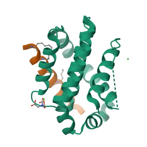
- The BAD gene encodes the Bcl2-associated agonist of cell death, also known as BBC6, BCL2L8, and Bcl-xL/Bcl-2-associated death promoter, a pro-apoptotic member of the Bcl-2 family.
- Bad promotes apoptosis by binding to and neutralizing anti-apoptotic proteins Bcl-X(L), Bcl-2, and Bcl-W, thereby freeing pro-apoptotic BAX to initiate mitochondrial outer membrane permeabilization.
- It acts as a critical signaling node linking growth factor receptor pathways to the apoptotic machinery; survival signals often suppress Bad via phosphorylation.
- Subcellular localization is dynamic: Bad resides in the cytoplasm and translocates to the mitochondrial outer membrane upon activation, where it exerts its pro-death function.
- Post-translational modifications including phosphorylation, acetylation, and methylation regulate Bad’s activity, stability, and interactions.
- Widely expressed across human tissues, Bad participates in homeostatic cell death regulation in diverse cell types.
Experimental Guidance and Technical Tips
- The immunogen, an N-terminal peptide (aa 1–100), suggests recognition of full‑length Bad; however, validation in your specific sample system is recommended to confirm reactivity with modified forms.
- In western blotting, a band at approximately 18 kDa is expected; use appropriate positive controls and consider phosphatase inhibitors if studying phosphorylation.
- For immunohistochemistry (paraffin), optimize antigen retrieval and antibody dilution for each tissue type; known positive tissues include lymphoid organs and many carcinomas.
- For ELISA applications, this antibody can detect recombinant or endogenous Bad; pairing with a distinct epitope‑specific antibody may improve sandwich assay performance.
- Due to Bad’s role as a phosphoprotein, include phosphatase inhibitors in lysis buffers when assessing activation status.
CamelBio: Your One-Stop Sourcing Bridge
CamelBio acts as your integrated sourcing partner for IVD raw materials, specializing in apoptosis and oncology targets like Bad. We supply validated antibody pairs, optimized monoclonal antibodies, and bulk ancillary reagents to accelerate diagnostic development. Contact us to streamline your project with comprehensive technical and supply‑chain support.
REQUEST A QUOTE
Our professional team will reply to you within one business day. Please feel free to contact us!